0
views
0
recommends
+1 Recommend
0 collections
    0
    shares
      • Record: found
      • Abstract: found
      • Article: found
      Is Open Access

      Corticocortical innervation subtypes of layer 5 intratelencephalic cells in the murine secondary motor cortex

      research-article

      Read this article at

      Bookmark
          There is no author summary for this article yet. Authors can add summaries to their articles on ScienceOpen to make them more accessible to a non-specialist audience.

          Abstract

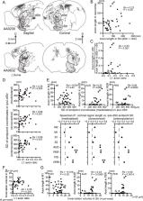
          Feedback projections from the secondary motor cortex (M2) to the primary motor and sensory cortices are essential for behavior selection and sensory perception. Intratelencephalic (IT) cells in layer 5 (L5) contribute feedback projections to diverse cortical areas. Here we show that L5 IT cells participating in feedback connections to layer 1 (L1) exhibit distinct projection patterns, genetic profiles, and electrophysiological properties relative to other L5 IT cells. An analysis of the MouseLight database found that L5 IT cells preferentially targeting L1 project broadly to more cortical regions, including the perirhinal and auditory cortices, and innervate a larger volume of striatum than the other L5 IT cells. We found experimentally that in upper L5 (L5a), ER81 (ETV1) was found more often in L1-preferring IT cells, and in IT cells projecting to perirhinal/auditory regions than those projecting to primary motor or somatosensory regions. The perirhinal region-projecting L5a IT cells were synaptically connected to each other and displayed lower input resistance than contra-M2 projecting IT cells including L1-preferring and nonpreferring cells. Our findings suggest that M2-L5a IT L1-preferring cells exhibit stronger ER81 expression and broader cortical/striatal projection fields than do cells that do not preferentially target L1.

          Related collections

          Most cited references66

          • Record: found
          • Abstract: found
          • Article: not found

          A robust and high-throughput Cre reporting and characterization system for the whole mouse brain

          The Cre/lox system is widely used in mice to achieve cell-type-specific gene expression. However, a strong and universal responding system to express genes under Cre control is still lacking. We have generated a set of Cre reporter mice with strong, ubiquitous expression of fluorescent proteins of different spectra. The robust native fluorescence of these reporters enables direct visualization of fine dendritic structures and axonal projections of the labeled neurons, which is useful in mapping neuronal circuitry, imaging and tracking specific cell populations in vivo. Using these reporters and a high-throughput in situ hybridization platform, we are systematically profiling Cre-directed gene expression throughout the mouse brain in a number of Cre-driver lines, including novel Cre lines targeting different cell types in the cortex. Our expression data are displayed in a public online database to help researchers assess the utility of various Cre-driver lines for cell-type-specific genetic manipulation.
            Bookmark
            • Record: found
            • Abstract: found
            • Article: not found

            Conserved cell types with divergent features in human versus mouse cortex

            Elucidating the cellular architecture of the human cerebral cortex is central to understanding our cognitive abilities and susceptibility to disease. Here we applied single nucleus RNA-sequencing to perform a comprehensive analysis of cell types in the middle temporal gyrus of human cortex. We identified a highly diverse set of excitatory and inhibitory neuronal types that are mostly sparse, with excitatory types being less layer-restricted than expected. Comparison to similar mouse cortex single cell RNA-sequencing datasets revealed a surprisingly well-conserved cellular architecture that enables matching of homologous types and predictions of human cell type properties. Despite this general conservation, we also find extensive differences between homologous human and mouse cell types, including dramatic alterations in proportions, laminar distributions, gene expression, and morphology. These species-specific features emphasize the importance of directly studying human brain.
              Bookmark
              • Record: found
              • Abstract: found
              • Article: not found

              Shared and distinct transcriptomic cell types across neocortical areas

              The neocortex contains a multitude of cell types that are segregated into layers and functionally distinct areas. To investigate the diversity of cell types across the mouse neocortex, here we analysed 23,822 cells from two areas at distant poles of the mouse neocortex: the primary visual cortex and the anterior lateral motor cortex. We define 133 transcriptomic cell types by deep, single-cell RNA sequencing. Nearly all types of GABA (γ-aminobutyric acid)-containing neurons are shared across both areas, whereas most types of glutamatergic neurons were found in one of the two areas. By combining single-cell RNA sequencing and retrograde labelling, we match transcriptomic types of glutamatergic neurons to their long-range projection specificity. Our study establishes a combined transcriptomic and projectional taxonomy of cortical cell types from functionally distinct areas of the adult mouse cortex.
                Bookmark

                Author and article information

                Contributors
                Journal
                Cereb Cortex
                Cereb Cortex
                cercor
                Cerebral Cortex (New York, NY)
                Oxford University Press
                1047-3211
                1460-2199
                01 January 2023
                09 April 2022
                09 April 2022
                : 33
                : 1
                : 50-67
                Affiliations
                National Institute for Physiological Sciences (NIPS) , Okazaki 444-8787, Japan
                Department of Physiological Sciences , The Graduate University for Advanced Studies (SOKENDAI) , Okazaki 444-8787, Japan
                Brain Science Institute , Tamagawa University, Machida, Tokyo 194-8610, Japan
                Department of Physiology , Division of Neurophysiology, School of Medicine, Tokyo Women's Medical University , Tokyo 162-8666, Japan
                National Institute for Physiological Sciences (NIPS) , Okazaki 444-8787, Japan
                Department of Physiological Sciences , The Graduate University for Advanced Studies (SOKENDAI) , Okazaki 444-8787, Japan
                National Institute for Physiological Sciences (NIPS) , Okazaki 444-8787, Japan
                Institute of Clinical Medicine and Research , Jikei University School of Medicine , Chiba 277-8567, Japan
                National Institute for Physiological Sciences (NIPS) , Okazaki 444-8787, Japan
                Department of Animal Physiology , Faculty of Veterinary Medicine, South Valley University , Qena 83523, Egypt
                Laboratory of Histology and Cytology, Faculty of Medicine, Hokkaido University , Sapporo 060-8638, Japan
                Section of Viral Vector Development , National Institute for Physiological Sciences , Okazaki 444-8585, Japan
                Bioresource Center, Gunma University Graduate School of Medicine , Gunma 371-8511, Japan
                KOKORO-Biology Group , Laboratories for Integrated Biology, Graduate School of Frontier Biosciences, Osaka University , Osaka 565-0871, Japan
                Physical and Health Education , Graduate School of Education, The University of Tokyo , Tokyo 113-0033, Japan
                International Research Center for Neurointelligence (WPI-IRCN), The University of Tokyo , Tokyo 113-0033, Japan
                National Institute for Physiological Sciences (NIPS) , Okazaki 444-8787, Japan
                Department of Physiological Sciences , The Graduate University for Advanced Studies (SOKENDAI) , Okazaki 444-8787, Japan
                Brain Science Institute , Tamagawa University, Machida, Tokyo 194-8610, Japan
                Author notes
                Corresponding author: Brain Science Institute, Tamagawa University Machida, Tokyo 1948610, Japan. Email: e191574@ 123456eve.tamagawa.ac.jp
                Author information
                https://orcid.org/0000-0003-3416-4903
                https://orcid.org/0000-0002-3073-9899
                https://orcid.org/0000-0002-7483-8362
                Article
                bhac052
                10.1093/cercor/bhac052
                9758586
                35396593
                c128fe97-545c-48bd-8d6c-f2ce5a086dba
                © The Author(s) 2022. Published by Oxford University Press on behalf of The Institute of Mathematics and its Applications. All rights reserved. For permissions, please email: journals.permissions@oup.com

                This is an Open Access article distributed under the terms of the Creative Commons Attribution-NonCommercial License ( https://creativecommons.org/licenses/by-nc/4.0/), which permits non-commercial re-use, distribution, and reproduction in any medium, provided the original work is properly cited. For commercial re-use, please contact journals.permissions@oup.com

                History
                : 15 March 2021
                : 25 January 2022
                : 26 January 2022
                Page count
                Pages: 18
                Funding
                Funded by: Ministry of Education, Culture, Sports, Science, and Technology;
                Award ID: 17H06311
                Award ID: 20H03359
                Funded by: Japan Society for the Promotion of Science, DOI 10.13039/501100001691;
                Award ID: 20H05049
                Categories
                Original Article
                AcademicSubjects/MED00310
                AcademicSubjects/MED00385

                Neurology
                intratelencephalic pyramidal cell,corticocortical,corticostriatal,mouselight,er81
                Neurology
                intratelencephalic pyramidal cell, corticocortical, corticostriatal, mouselight, er81

                Comments

                Comment on this article