74
views
0
recommends
+1 Recommend
0 collections
    0
    shares
      • Record: found
      • Abstract: found
      • Article: found
      Is Open Access

      Small incision lenticule extraction (SMILE) history, fundamentals of a new refractive surgery technique and clinical outcomes

      review-article

      Read this article at

      Bookmark
          There is no author summary for this article yet. Authors can add summaries to their articles on ScienceOpen to make them more accessible to a non-specialist audience.

          Abstract

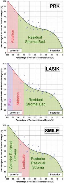
          This review summarizes the current status of the small incision lenticule extraction (SMILE) procedure. Following the early work by Sekundo et al. and Shah et al., SMILE has become increasingly popular. The accuracy of the creation of the lenticule with the VisuMax femtosecond laser (Carl Zeiss Meditec) has been verified using very high-frequency (VHF) digital ultrasound and optical coherence tomography (OCT). Visual and refractive outcomes have been shown to be similar to those achieved with laser in situ keratomileusis (LASIK), notably in a large population reported by Hjortdal, Vestergaard et al. Safety in terms of the change in corrected distance visual acuity (CDVA) has also been shown to be similar to LASIK. It was expected that there would be less postoperative dry eye after SMILE compared to LASIK because the anterior stroma is disturbed only by the small incision, meaning that the anterior corneal nerves should be less affected. A number of studies have demonstrated a lower reduction and faster recovery of corneal sensation after SMILE than LASIK. Some studies have also used confocal microscopy to demonstrate a lower decrease in subbasal nerve fiber density after SMILE than LASIK. The potential biomechanical advantages of SMILE have been modeled by Reinstein et al. based on the non-linearity of tensile strength through the stroma. Studies have reported a similar change in Ocular Response Analyzer (Reichert) parameters after SMILE and LASIK, however, these have previously been shown to be unreliable as a representation of corneal biomechanics. Retreatment options after SMILE are discussed. Tissue addition applications of the SMILE procedure are also discussed including the potential for cryo-preservation of the lenticule for later reimplantation (Mohamed-Noriega, Angunawela, Lim et al.), and a new procedure referred to as endokeratophakia in which a myopic SMILE lenticule is implanted into a hyperopic patient (Pradhan et al.). Finally, studies reporting microdistortions in Bowman’s layer and corneal wound healing responses are also described.

          Additional non-English abstract (French, Francais)

          Cette revue résume les connaissances actuelles sur la procedure SMILE (small incision lenticule extraction). Apres les travaux préliminaires des groupes de Sekundo et al. et de Shah et al., la procedure SMILE est désormais devenue courante. Le laser femtosecond VisuMax (Carl Zeiss Meditec) permet une découpe précise du lenticule, comme cela a été vérifié par ultrasound numérique à tres haute fréquence (Artemis) et par OCT. Il a été démontré, entre autres par Hjortdal, Vestergaard, et al. dans leur etude sur une population large, que les résultats visuels et refractifs sont comparables à ceux du LASIK, et que le risque de perte de lignes d’acuité corrigée est aussi semblable à celui du LASIK. Avec SMILE, contrairement au LASIK, la partie antérieure du stroma étant seulement découpée par une incision de petite taille, les nerfs cornéens antérieurs sont moins touchés et par consequent, le taux d’yeux secs en phase post-opératoire devrait etre plus faible après SMILE qu’apres LASIK. Quelques études ont effectivement démontré que la sensation cornéenne était moins réduite après SMILE qu’après LASIK et que la récupération de la sensation cornéenne était plus rapide. Quelques études ont aussi utilisé la microscopie confocale pour prouver qu’il y avait une diminution plus faible de la densité des fibres nerveuses de la couche sub-basale après SMILE qu’apres LASIK. Au niveau biomécanique, Reinstein et al. ont proposé un modèle basé sur la non-linéarité de la force de tension du stroma, mettant en avant les avantages potentiels de SMILE. Certaines études ont comparé les paramètres mesurés par l’Ocular Response Analayzer (Reichert) et rapporté un changement similaire après SMILE ou LASIK; cependant, on sait que ces paramètres ne peuvent pas etre pris en compte pour représenter la biomécanique cornéenne. Les options de re-traitement après SMILE sont présentées, ainsi que la possibilité d’ajouter du tisuu cornéen: par example, le potential de cryo-preservation du lenticule pour une ré-implantation ultérieure (Mohamed-Noriega, Angunawela, Lim et al.), et une nouvelle procedure nommée endokératophakie dans laquelle un lenticule SMILE est implanté dans la cornée d’un oeil hypermétrope (Pradhan et al.). Pour finir, des études rapportant des micro-distortions dans la couche de Bowmans et des études sur le processus de reparation de la cornée sont décrites.

          Related collections

          Most cited references83

          • Record: found
          • Abstract: found
          • Article: not found

          Results of small incision lenticule extraction: All-in-one femtosecond laser refractive surgery.

          To report the clinical results of small incision lenticule extraction to correct refractive errors using a femtosecond laser to refine the femtosecond lenticule extraction technique. Private laser center, Vadodara, India. Prospective clinical study. The VisuMax femtosecond laser system was used to perform small incision lenticule extraction to treat refractive errors. The laser was used to cut a refractive lenticule intrastromally to correct myopia and myopic astigmatism. The lenticule was then extracted from the stroma through a 3.0 to 5.0 mm incision. Outcome measures were corrected distance visual acuity (CDVA), uncorrected distance visual acuity (UDVA), and manifest refraction during 6 months of follow-up. Corneal topography and ocular wavefront aberrations were also measured. The study enrolled 51 eyes of 41 patients. The mean spherical equivalent was -4.87 diopters (D) ± 2.16 (SD) preoperatively and +0.03 ± 0.30 D 6 months postoperatively. Refractive stability was achieved within 1 month (P<.01). Six months after surgery, 79% of all full-correction cases had a UDVA of 20/25 or better. The 6-month postoperative CDVA was the same as or better than the preoperative CDVA in 95% of eyes. Two eyes lost 1 line of CDVA. All-in-one femtosecond refractive correction using a small incision technique was safe, predictable, and effective in treating myopia and myopic astigmatism. No author has a financial or proprietary interest in any material or method mentioned. Additional disclosure is found in the footnotes. Copyright © 2011 ASCRS and ESCRS. Published by Elsevier Inc. All rights reserved.
            Bookmark
            • Record: found
            • Abstract: found
            • Article: not found

            Safety and complications of more than 1500 small-incision lenticule extraction procedures.

            To evaluate the safety and complications of small-incision lenticule extraction (SMILE). Clinical control cohort study. A total of 922 healthy patients (1800 eyes) who were treated for myopia or myopic astigmatism between January 2011 and March 2013 at the Department of Ophthalmology, Aarhus, Denmark. Patients received a full preoperative examination and were treated with SMILE in both eyes and followed for 3 months (1574 eyes). Patients with complications, including loss of corrected distance visual acuity (CDVA) or dissatisfaction, were offered a late reexamination. Surgical complications and CDVA. Mean preoperative spherical equivalent refraction was -7.25±1.84 diopters (D). Average postoperative refraction was -0.28±0.52 D, and mean error of treatment was -0.15±0.50 D. By 3 months, 86% (1346 eyes) had unchanged or improved CDVA. A loss of 2 or more lines was observed in 1.5% of eyes; however, at a late follow-up visit (average, 18 months), CDVA was within 1 line of the preoperative level in all eyes. Perioperative complications included epithelial abrasions (6%), small tears at the incision (1.8%), and difficult lenticule extraction (1.9%). The cap was perforated in 4 eyes, and a major tear occurred in 1 eye; however, none of these patients had late visual symptoms. In 0.8% (14 eyes), suction was lost during surgery. Re-treatment was successful in 13 eyes, whereas 1 eye had ghost images and was re-treated with topography-guided photorefractive keratectomy (PRK). Postoperative complications included trace haze (8%), epithelial dryness on day 1 (5%), interface inflammation secondary to central abrasion (0.3%), and minor interface infiltrates (0.3%); these complications had an impact on CDVA at 3 months in only 1 case. Irregular corneal topography occurred in 1.0% of eyes, resulting in reduced 3-month CDVA (12 eyes) or ghost images (6 eyes). Topography-guided PRK was performed in 4 eyes, with improvement in 3 cases. Satisfaction was high, with only 2 patients dissatisfied at their latest visit because of blurred vision or residual astigmatism. Overall, SMILE had acceptable safety. Although 1.5% of eyes had reduced CDVA by 3 months, visual acuity was restored in the long term. Likewise, patient satisfaction was high. Copyright © 2014 American Academy of Ophthalmology. Published by Elsevier Inc. All rights reserved.
              Bookmark
              • Record: found
              • Abstract: found
              • Article: not found

              Biomechanical evidence of the distribution of cross-links in corneas treated with riboflavin and ultraviolet A light.

              To examine to which depth of the cornea the stiffening effect is biomechanically detectable. Department of Ophthalmology, University of Dresden, Dresden, Germany. Of 40 enucleated porcine eyes, 20 eyes were treated with the photosensitizer riboflavin (0.1%) and ultraviolet A (UVA) light (370 nm, 3 mW/cm2, 30 minutes); the other 20 eyes served as control. From each eye, 2 flaps of 200 microm thickness were cut with a microkeratome, and strips of 5 mm width and 7 mm length were prepared. Stress-strain behavior was measured with a material tester to characterize the stiffening effect. Five pairs of human donor eyes were tested in the same way. In porcine corneas, the stiffening effect was stronger in the anterior-treated flaps than in the posterior-treated flaps and the control flaps (P = .001). A 5% strain was achieved at a stress of 261.7 +/- 133.2 x 10(3) N/m2 in the anterior-treated flaps and 104.1 +/- 52.7 x 10(3) N/m2 in the anterior control flaps. The posterior-treated flaps (105.0 +/- 55.8 x 10(3) N/m2) and the posterior control flaps (103.7 +/- 61.8 x 10(3) N/m2) showed no difference (P = .95). A similar stiffening effect was observed in human eyes, but contrary to findings in porcine corneas, in human corneas the anterior control flaps were stiffer than the posterior control flaps (P = .027). Treatment of the cornea with riboflavin and UVA significantly stiffened the cornea only in the anterior 200 microm. This depth-dependent stiffening effect may be explained by the absorption behavior for UVA in the riboflavin-treated cornea. Sixty-five percent to 70% of UVA irradiation was absorbed within the anterior 200 microm and only 20% in the next 200 microm. Therefore, deeper structures and even the endothelium are not affected.
                Bookmark

                Author and article information

                Contributors
                dzr@londonvisionclinic.com
                tim@londonvisionclinic.com
                marine@londonvisionclinic.com
                Journal
                Eye Vis (Lond)
                Eye Vis (Lond)
                Eye and Vision
                BioMed Central (London )
                2326-0254
                16 October 2014
                16 October 2014
                2014
                : 1
                : 3
                Affiliations
                [ ]London Vision Clinic, 138 Harley Street, London, W1G 7LA UK
                [ ]Department of Ophthalmology, Columbia University Medical Center, New York, NY USA
                [ ]Centre Hospitalier National d’Ophtalmologie, Paris, France
                Article
                3
                10.1186/s40662-014-0003-1
                4604118
                26605350
                c05f4dba-5762-4ea4-a33d-28fbaf05124e
                © Reinstein et al.; licensee BioMed Central 2014

                This is an Open Access article distributed under the terms of the Creative Commons Attribution License ( http://creativecommons.org/licenses/by/4.0), which permits unrestricted use, distribution, and reproduction in any medium, provided the original work is properly credited. The Creative Commons Public Domain Dedication waiver ( http://creativecommons.org/publicdomain/zero/1.0/) applies to the data made available in this article, unless otherwise stated.

                History
                : 19 May 2014
                : 13 August 2014
                Categories
                Review
                Custom metadata
                © The Author(s) 2014

                small incision lenticule extraction (smile),laser in situ keratomileusis (lasik),lenticule,cap,dry eye,corneal sensation,corneal innervation,corneal biomechanics,endokeratophakia

                Comments

                Comment on this article