17
views
0
recommends
+1 Recommend
0 collections
    0
    shares
      • Record: found
      • Abstract: found
      • Article: found
      Is Open Access

      Comparative analysis of tools for live cell imaging of actin network architecture

      research-article

      Read this article at

      Bookmark
          There is no author summary for this article yet. Authors can add summaries to their articles on ScienceOpen to make them more accessible to a non-specialist audience.

          Abstract

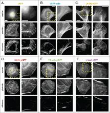
          Fluorescent derivatives of actin and actin-binding domains are powerful tools for studying actin filament architecture and dynamics in live cells. Growing evidence, however, indicates that these probes are biased, and their cellular distribution does not accurately reflect that of the cytoskeleton. To understand the strengths and weaknesses of commonly used live-cell probes—fluorescent protein fusions of actin, Lifeact, F-tractin, and actin-binding domains from utrophin—we compared their distributions in cells derived from various model organisms. We focused on five actin networks: the peripheral cortex, lamellipodial and lamellar networks, filopodial bundles, and stress fibers. Using phalloidin as a standard, we identified consistent biases in the distribution of each probe. The localization of F-tractin is the most similar to that of phalloidin but induces organism-specific changes in cell morphology. Both Lifeact and GFP-actin concentrate in lamellipodial actin networks but are excluded from lamellar networks and filopodia. In contrast, the full utrophin actin-binding domain (Utr261) binds filaments of the lamellum but only weakly localizes to lamellipodia, while a shorter variant (Utr230) is restricted to the most stable subpopulations of actin filaments: cortical networks and stress fibers. In some cells, Utr230 also detects Golgi-associated filaments, previously detected by immunofluorescence but not visible by phalloidin staining. Consistent with its localization, Utr230 exhibits slow rates of fluorescence recovery after photobleaching (FRAP) compared to F-tractin, Utr261 and Lifeact, suggesting that it may be more useful for FRAP- and photo-activation-based studies of actin network dynamics.

          Related collections

          Most cited references15

          • Record: found
          • Abstract: found
          • Article: not found

          Two distinct actin networks drive the protrusion of migrating cells.

          Cell migration initiates by extension of the actin cytoskeleton at the leading edge. Computational analysis of fluorescent speckle microscopy movies of migrating epithelial cells revealed this process is mediated by two spatially colocalized but kinematically, kinetically, molecularly, and functionally distinct actin networks. A lamellipodium network assembled at the leading edge but completely disassembled within 1 to 3 micrometers. It was weakly coupled to the rest of the cytoskeleton and promoted the random protrusion and retraction of the leading edge. Productive cell advance was a function of the second colocalized network, the lamella, where actomyosin contraction was integrated with substrate adhesion.
            Bookmark
            • Record: found
            • Abstract: found
            • Article: not found

            Versatile fluorescent probes for actin filaments based on the actin-binding domain of utrophin.

            Actin filaments (F-actin) are protein polymers that undergo rapid assembly and disassembly and control an enormous variety of cellular processes ranging from force production to regulation of signal transduction. Consequently, imaging of F-actin has become an increasingly important goal for biologists seeking to understand how cells and tissues function. However, most of the available means for imaging F-actin in living cells suffer from one or more biological or experimental shortcomings. Here we describe fluorescent F-actin probes based on the calponin homology domain of utrophin (Utr-CH), which binds F-actin without stabilizing it in vitro. We show that these probes faithfully report the distribution of F-actin in living and fixed cells, distinguish between stable and dynamic F-actin, and have no obvious effects on processes that depend critically on the balance of actin assembly and disassembly. (c) 2007 Wiley-Liss, Inc.
              Bookmark
              • Record: found
              • Abstract: found
              • Article: not found

              Cofilin Changes the Twist of F-Actin: Implications for Actin Filament Dynamics and Cellular Function

              Cofilin is an actin depolymerizing protein found widely distributed in animals and plants. We have used electron cryomicroscopy and helical reconstruction to identify its binding site on actin filaments. Cofilin binds filamentous (F)-actin cooperatively by bridging two longitudinally associated actin subunits. The binding site is centered axially at subdomain 2 of the lower actin subunit and radially at the cleft between subdomains 1 and 3 of the upper actin subunit. Our work has revealed a totally unexpected (and unique) property of cofilin, namely, its ability to change filament twist. As a consequence of this change in twist, filaments decorated with cofilin have much shorter ‘actin crossovers' (∼75% of those normally observed in F-actin structures). Although their binding sites are distinct, cofilin and phalloidin do not bind simultaneously to F-actin. This is the first demonstration of a protein that excludes another actin-binding molecule by changing filament twist. Alteration of F-actin structure by cofilin/ADF appears to be a novel mechanism through which the actin cytoskeleton may be regulated or remodeled.
                Bookmark

                Author and article information

                Journal
                Bioarchitecture
                Bioarchitecture
                KBIA
                Bioarchitecture
                Taylor & Francis
                1949-0992
                1949-100X
                2014
                28 August 2015
                28 August 2015
                : 4
                : 6
                : 189-202
                Affiliations
                Cellular and Molecular Pharmacology; University of California ; San Francisco, CA USA
                Author notes
                [* ]Address correspondence to: R Dyche Mullins; Email: dyche@ 123456mullinslab.ucsf.edu
                Article
                1047714
                10.1080/19490992.2014.1047714
                4914014
                26317264
                bfa4db59-14c2-4a0a-910b-f6ac00416558
                © 2015 The Author(s). Published with license by Taylor & Francis Group, LLC

                This is an Open Access article distributed under the terms of the Creative Commons Attribution-Non-Commercial License http://creativecommons.org/licenses/by-nc/3.0/, which permits unrestricted non-commercial use, distribution, and reproduction in any medium, provided the original work is properly cited. The moral rights of the named author(s) have been asserted.

                History
                : 28 March 2015
                : 8 June 2015
                : 31 March 2015
                Page count
                Figures: 8, Tables: 3, References: 20, Pages: 14
                Categories
                Research Paper

                Molecular biology
                cytoskeleton,actin,cell architecture,live cell imaging,fluorescent protein reporters

                Comments

                Comment on this article