15
views
0
recommends
+1 Recommend
0 collections
    0
    shares
      • Record: found
      • Abstract: found
      • Article: found
      Is Open Access

      Bone tissue regeneration: The role of finely tuned pore architecture of bioactive scaffolds before clinical translation

      research-article

      Read this article at

      Bookmark
          There is no author summary for this article yet. Authors can add summaries to their articles on ScienceOpen to make them more accessible to a non-specialist audience.

          Abstract

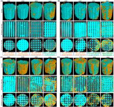
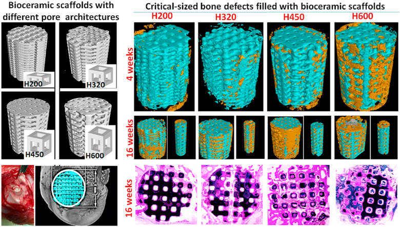
          Spatial dimension of pores and interconnection in macroporous scaffolds is of particular importance in facilitating endogenous cell migration and bone tissue ingrowth. However, it is still a challenge to widely tune structure parameters of scaffolds by conventional methods because of inevitable pore geometrical deformation and poor pore interconnectivity. Here, the long-term in vivo biological performances of nonstoichiometric bioceramic scaffolds with different pore dimensions were assessed in critical-size femoral bone defect model. The 6% Mg-substituted wollastonite (CSi-Mg6) powders were prepared via wet-chemical precipitation and the scaffolds elaborately printed by ceramic stereolithography, displaying designed constant pore strut and tailorable pore height (200, 320, 450, 600 μm), were investigated thoroughly in the bone regeneration process. Together with detailed structural stability and mechanical properties were collaboratively outlined. Both μCT and histological analyses indicated that bone tissue ingrowth was retarded in 200 μm scaffolds in the whole stage (2–16 weeks) but the 320 μm scaffolds showed appreciable bone tissue in the center of porous constructs at 6–10 weeks and matured bone tissue were uniformly invaded in the whole pore networks at 16 weeks. Interestingly, the neo-tissue ingrowth was facilitated in the 450 μm and 600 μm scaffolds after 2 weeks and higher extent of bone regeneration and remodeling at the later stage. These new findings provide critical information on how engineered porous architecture impact bone regeneration in vivo. Simultaneously, this study shows important implications for optimizing the porous scaffolds design by advanced additive manufacture technique to match the clinical translation with high performance.

          Graphical abstract

          Highlights

          • 6% Mg-substituted wollastonite (CSi-Mg6) bioceramic show appreciable bioactivity and mechanical strength.

          • Porous CSi-Mg6 scaffolds with precisely controlled pore dimensions are fabricated by ceramic stereolithography.

          • The favorable pore geometries facilitating neo-bone ingrowth into the center pores of scaffolds are decoded.

          • CAD-assisted stereolithography opens up opportunities for developing scaffolds with tailored pore architecture.

          Related collections

          Most cited references59

          • Record: found
          • Abstract: found
          • Article: not found

          Porosity of 3D biomaterial scaffolds and osteogenesis.

          Porosity and pore size of biomaterial scaffolds play a critical role in bone formation in vitro and in vivo. This review explores the state of knowledge regarding the relationship between porosity and pore size of biomaterials used for bone regeneration. The effect of these morphological features on osteogenesis in vitro and in vivo, as well as relationships to mechanical properties of the scaffolds, are addressed. In vitro, lower porosity stimulates osteogenesis by suppressing cell proliferation and forcing cell aggregation. In contrast, in vivo, higher porosity and pore size result in greater bone ingrowth, a conclusion that is supported by the absence of reports that show enhanced osteogenic outcomes for scaffolds with low void volumes. However, this trend results in diminished mechanical properties, thereby setting an upper functional limit for pore size and porosity. Thus, a balance must be reached depending on the repair, rate of remodeling and rate of degradation of the scaffold material. Based on early studies, the minimum requirement for pore size is considered to be approximately 100 microm due to cell size, migration requirements and transport. However, pore sizes >300 microm are recommended, due to enhanced new bone formation and the formation of capillaries. Because of vascularization, pore size has been shown to affect the progression of osteogenesis. Small pores favored hypoxic conditions and induced osteochondral formation before osteogenesis, while large pores, that are well-vascularized, lead to direct osteogenesis (without preceding cartilage formation). Gradients in pore sizes are recommended for future studies focused on the formation of multiple tissues and tissue interfaces. New fabrication techniques, such as solid-free form fabrication, can potentially be used to generate scaffolds with morphological and mechanical properties more selectively designed to meet the specificity of bone-repair needs.
            Bookmark
            • Record: found
            • Abstract: not found
            • Article: not found

            Biomaterials & scaffolds for tissue engineering

              Bookmark
              • Record: found
              • Abstract: found
              • Article: found
              Is Open Access

              Bone grafts and biomaterials substitutes for bone defect repair: A review

              Bone grafts have been predominated used to treat bone defects, delayed union or non-union, and spinal fusion in orthopaedic clinically for a period of time, despite the emergency of synthetic bone graft substitutes. Nevertheless, the integration of allogeneic grafts and synthetic substitutes with host bone was found jeopardized in long-term follow-up studies. Hence, the enhancement of osteointegration of these grafts and substitutes with host bone is considerably important. To address this problem, addition of various growth factors, such as bone morphogenetic proteins (BMPs), parathyroid hormone (PTH) and platelet rich plasma (PRP), into structural allografts and synthetic substitutes have been considered. Although clinical applications of these factors have exhibited good bone formation, their further application was limited due to high cost and potential adverse side effects. Alternatively, bioinorganic ions such as magnesium, strontium and zinc are considered as alternative of osteogenic biological factors. Hence, this paper aims to review the currently available bone grafts and bone substitutes as well as the biological and bio-inorganic factors for the treatments of bone defect.
                Bookmark

                Author and article information

                Contributors
                Journal
                Bioact Mater
                Bioact Mater
                Bioactive Materials
                KeAi Publishing
                2452-199X
                07 November 2020
                May 2021
                07 November 2020
                : 6
                : 5
                : 1242-1254
                Affiliations
                [a ]Department of Orthopedics, The First Affiliated Hospital, Zhejiang University School of Medicine, Hangzhou, 310003, China
                [b ]Bio-nanomaterials and Regenerative Medicine Research Division, Zhejiang-California International Nanosystem Institute, Zhejiang University, Hangzhou, 310058, China
                [c ]Rui'an People's Hospital & the 3rd Hospital Affiliated to Wenzhou Medical University, Rui’ An, 325200, China
                Author notes
                []Corresponding author. Zhejiang-California International Nanosystems Institute Zhejiang University, Hangzhou, Zhejiang Province, 310058, China zhrgou@ 123456zju.edu.cn
                [∗∗ ]Corresponding author. Department of Orthopedics, the First Affiliated Hospital Zhejiang University School of Medicine 79# Qingchun Road, Hangzhou, Zhejiang Province, 310003, China xusanzhong@ 123456zju.edu.cn
                [1]

                These authors contributed equally.

                Article
                S2452-199X(20)30291-7
                10.1016/j.bioactmat.2020.11.003
                7653208
                33210022
                bdd80b4b-7d5f-48dc-a393-fcc4439c04b8
                © 2020 [The Author/The Authors]

                This is an open access article under the CC BY-NC-ND license (http://creativecommons.org/licenses/by-nc-nd/4.0/).

                History
                : 21 September 2020
                : 31 October 2020
                : 1 November 2020
                Categories
                Article

                pore structural parameter,bone regeneration efficiency,precise manufacturing,porous scaffolds,tissue engineering

                Comments

                Comment on this article