12
views
0
recommends
+1 Recommend
0 collections
    0
    shares
      • Record: found
      • Abstract: found
      • Article: found
      Is Open Access

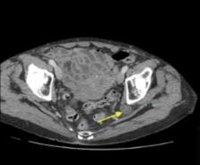
      Ureterosciatic Hernia in Focus: A Narrative Review of the Literature

      review-article
      1 , , 1 , 1 , 1 , 1
      ,
      Cureus
      Cureus
      obstructive uropathy, sciatic ureteral hernia, curlicue sign, lindbom hernia, uretero-sciatic hernia

      Read this article at

      Bookmark
          There is no author summary for this article yet. Authors can add summaries to their articles on ScienceOpen to make them more accessible to a non-specialist audience.

          Abstract

          Pelvic herniation of the ureter through anatomical musculoskeletal foramina stands out as one of the rarest causes of ureteric obstruction. Historically, most cases have been documented as incidental intraoperative findings. The herniation of the ureter through the sciatic foramen presents as a particularly uncommon variant of this condition, distinguished by its potential to cause life-threatening sepsis or renal failure if not promptly recognized and treated. The diagnostic process remains challenging, attributed partly to the vague initial symptomatology and subtle radiological findings, and second, to the rarity of this condition. This challenge may be further compounded by the lack of a clear description of clinical features and pathways to raise clinician suspicion. In light of these considerations, we conducted this literature review to illuminate this unique cause of obstructive uropathy, aiming to delineate its clinical features and explore common diagnostic and treatment options.

          Related collections

          Most cited references50

          • Record: found
          • Abstract: found
          • Article: not found

          Unenhanced helical computed tomography vs intravenous urography in patients with acute flank pain: accuracy and economic impact in a randomized prospective trial.

          Unenhanced helical computed tomography (UHCT) has evolved into a well-accepted alternative to intravenous urography (IVU) in patients with acute flank pain and suspected ureterolithiasis. The purpose of our randomized prospective study was to analyse the diagnostic accuracy of UHCT vs IVU in the normal clinical setting with special interest on economic impact, applied radiation dose and time savings in patient management. A total of 122 consecutive patients with acute flank pain suggestive of urolithiasis were randomized for UHCT ( n=59) or IVU ( n=63). Patient management (time, contrast media), costs and radiation dose were analysed. The films were independently interpreted by four radiologists, unaware of previous findings, clinical history and clinical outcome. Alternative diagnoses if present were assessed. Direct costs of UHCT and IVU are nearly identical (310/309 Euro). Indirect costs are much lower for UHCT because it saves examination time and when performed immediately initial abdominal plain film (KUB) and sonography are not necessary. Time delay between access to the emergency room and start of the imaging procedure was 32 h 7 min for UHCT and 36 h 55 min for IVU. The UHCT took an average in-room time of 23 min vs 1 h 21 min for IVU. Mild to moderate adverse reactions for contrast material were seen in 3 (5%) patients. The UHCT was safe, as no contrast material was needed. The mean applied radiation dose was 3.3 mSv for IVU and 6.5 mSv for UHCT. Alternative diagnoses were identified in 4 (7%) UHCT patients and 3 (5%) IVU patients. Sensitivity and specificity of UHCT and IVU was 94.1 and 94.2%, and 85.2 and 90.4%, respectively. In patients with suspected renal colic KUB and US may be the least expensive and most easily accessable modalities; however, if needed and available, UHCT can be considered a better alternative than IVU because it has a higher diagnostic accuracy and a better economic impact since it is more effective, faster, less expensive and less risky than IVU. In addition, it also has the capability of detecting various additional renal and extrarenal pathologies.
            Bookmark
            • Record: found
            • Abstract: found
            • Article: not found

            Ureterosciatic Hernia: A Rare Cause of Ureteral Obstruction Visualized by Multislice Helical Computed Tomography

            Ureterosciatic herniation is an extremely rare cause of ureteral obstruction, of which few cases have been published. We describe a case revealed by pyelonephritis with acute renal failure in an 81-year-old woman. After percutaneous nephrostomy tube placement and antibiotic therapy, urography and multiplanar computed tomography reconstructions of the pelvis confirmed the diagnosis. The symptoms resolved, and the hernia was then corrected surgically.
              Bookmark
              • Record: found
              • Abstract: found
              • Article: not found

              Laparoscopic repair of ureterosciatic hernia.

              Ureterosciatic herniation is a rare benign event that can mimic diverticulosis or irritable bowel syndrome. This entity has been managed by a number of open surgical techniques. Laparoscopic repair of this entity enabled us to identify the defect, interpose mesh, and obliterate the hernia defect with minimal morbidity. This represents the first report of laparoscopic repair of a ureterosciatic hernia.
                Bookmark

                Author and article information

                Journal
                Cureus
                Cureus
                2168-8184
                Cureus
                Cureus (Palo Alto (CA) )
                2168-8184
                4 December 2023
                December 2023
                : 15
                : 12
                : e49895
                Affiliations
                [1 ] Urology, James Paget University Hospitals NHS Foundation Trust, Great Yarmouth, GBR
                Author notes
                Article
                10.7759/cureus.49895
                10762697
                38174201
                b9c796fe-a76c-4c8f-81f1-fa1ede65bf67
                Copyright © 2023, Mustafa et al.

                This is an open access article distributed under the terms of the Creative Commons Attribution License CC-BY 4.0., which permits unrestricted use, distribution, and reproduction in any medium, provided the original author and source are credited.

                History
                : 4 December 2023
                Categories
                Urology

                obstructive uropathy,sciatic ureteral hernia,curlicue sign,lindbom hernia,uretero-sciatic hernia

                Comments

                Comment on this article