15
views
0
recommends
+1 Recommend
0 collections
    0
    shares
      • Record: found
      • Abstract: found
      • Article: found
      Is Open Access

      Intralesional corticosteroid injections in the treatment of central giant cell lesions of the jaws: A meta-analytic study

      review-article

      Read this article at

      Bookmark
          There is no author summary for this article yet. Authors can add summaries to their articles on ScienceOpen to make them more accessible to a non-specialist audience.

          Abstract

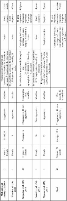
          Objective: The aim of this study was to evaluate the response of treatment of central giant cell lesion to intralesional corticosteroid injections. Study Design: Review of articles indexed in PubMed on the topic between the years 1988 and 2011, and development of a descriptive meta-analysis of the results. Results: Sample of 41 patients primarily treated with intralesional corticosteroid injections was obtained, with a male female ratio of 1:0.95, being 23 aggressive and 18 non-aggressive central giant cell lesions. Triamcinolone acetonide and triamcinolone hexacetonide were the drugs used, and 78.0% cases were considered as good result, 14.6% were considered as moderate response and 7.3% were considered as negative result to treatment. Considering the aggressiveness, 88.9% of non-aggressive lesions presented a good response to treatment, in aggressive central giant cell lesions, 69.6% presented a good response to intralesional corticosteroid injections. Conclusion: In view of the results analyzed, intralesional corticosteroid injections could be considered as first treatment option for central giant cell lesion.

          Key words:Central giant cell lesion, corticosteroids injections, triamcinolone hexacetonide, triamcinolone acetonide.

          Related collections

          Most cited references35

          • Record: found
          • Abstract: found
          • Article: not found

          Central giant cell lesions of the jaws: a clinicopathologic study.

          The biologic behavior of central giant cell lesions of the jaws ranges from quiescent to aggressive with destructive expansion. To date, these variations have not been explained by the findings of routine histologic examination. This retrospective clinicopathologic study of giant cell lesions was performed to search for histologic correlates of biologic behavior. Lesions in 17 patients were classified clinically as nonaggressive (group I) or aggressive (group II). In general, group II lesions affected children at an earlier age, were larger at the time of diagnosis, and recurred more frequently. The following histologic parameters were assessed: fractional surface area occupied by giant cells (FSA), relative size index of giant cells (RSI), stromal characteristics, mitotic index, inflammatory cells, and hemosiderin content. Histologic differences between the two groups were not as clear as the differences in biologic behavior. However, aggressive lesions had a higher RSI, and recurrent giant cells lesions had a higher RSI and FSA; these parameters warrant further study. In addition, electron microscopic differences in a small number of aggressive and nonaggressive lesions were documented.
            Bookmark
            • Record: found
            • Abstract: found
            • Article: not found

            Central giant cell granuloma of the jaws: a clinical, radiologic, and histopathologic study of 26 cases.

            The clinical behavior of central giant cell granuloma (CGCG) of the jaws is variable and difficult to predict. Clinical data and follow-up information of 26 patients with CGCG were analyzed. Histologic features were correlated with the clinical course of the disease. In 16 patients the CGCGs were asymptomatic; 10 lesions presented with aggressive growth, pain, massive swelling, root resorption, cortical perforation, and/or recurrence. These patients were younger and the lesions were larger than in the nonaggressive group. The histomorphometric analysis proved a significant increase in large giant cells, fractional surface area, and mitotic activity in aggressive CGCG lesions. Immunohistologic investigation (Ki-67 and p53 stain) revealed no significant differences. After surgical treatment, 3 patients with aggressive lesions developed a recurrence. The data show that clinical and histomorphometric features may be reliable indicators for the differentiation between aggressive and nonaggressive CGCG. This should be accounted for to improve the individual planning of the treatment and follow-up.
              Bookmark
              • Record: found
              • Abstract: found
              • Article: not found

              Clinical and radiological features of central giant-cell lesions of the jaw.

              The purpose of this study was to evaluate the clinical and radiological features of central giant-cell lesions that were diagnosed in The Netherlands between January 1, 1990, and January 1, 1995. A population-based retrospective study was carried out, examining all patients with a central giant-cell lesion from this period. In 83 patients there was a central giant-cell granuloma (89 lesions). Aggressive signs and symptoms (pain, paresthesia, or root resorption) were found in 16 (19.3%) patients. Multiple lesions occurred in 3 (3.6%) patients. The overall recurrence rate was 26.3%, and there was a higher recurrence rate in patients who exhibited aggressive signs and symptoms than in patients without these features (RR 1.6). In 5 patients a clinical diagnosis of cherubism or concomitant neurofibromatosis type 1 was made (14 lesions). In a general population, large and aggressive lesions are less common than suggested by the literature. Multiple lesions, however, occur more frequently than previously assumed. In patients with aggressive signs and symptoms, surgical curettage is not an effective therapy.
                Bookmark

                Author and article information

                Journal
                Med Oral Patol Oral Cir Bucal
                Med Oral Patol Oral Cir Bucal
                Medicina Oral S.L.
                Medicina Oral, Patología Oral y Cirugía Bucal
                Medicina Oral S.L.
                1698-4447
                1698-6946
                March 2013
                5 February 2013
                : 18
                : 2
                : e226-e232
                Affiliations
                [1 ]DDS, MSc, Assistant Professor, Department of Pathology, Fortaleza University School of Medicine, Fortaleza, Brazil
                [2 ]DDS, MSc student, Federal University of Ceará School of Dentistry, Fortaleza, Brazil
                [3 ]DDS, MSc, PhD, Assistant Professor, Faculdade Católica Rainha do Sertão, School of Dentistry, Quixadá, Brazil
                [4 ]DDS, MSc, PhD, Associate Professor, Department of Oral and Maxillofacial Pathology, Fortaleza University School of Dentistry, Fortaleza, Brazil
                [5 ]DDS, MSc, PhD, Associate Professor of Oral and Maxillofacial Surgery, Bauru School of Dentistry, University of São Paulo, Bauru, Brazil
                [6 ]DDS, MSc, PhD, Associate Professor, Department of Dental Clinic, Discipline of Oral and Maxillofacial Surgery and Stomatology, Federal University of Ceara School of Dentistry, Fortaleza, Brazil. Oral and Maxillofacial Surgeon, Department of Oral and Maxillofacial Surgery, Memorial Batista Hospital, Fortaleza, Brazil
                Author notes
                Universidade de Fortaleza Fortaleza University/ Universidad de Fortaleza Av. Washington Soares 1321, Edson Queiroz 60811-905 Fortaleza Ceará, Brasil P.O. Box 1258 , E-mail: rlimaverde@ 123456unifor.br
                Article
                18345
                10.4317/medoral.18345
                3613330
                23385503
                b94fffb8-31b6-4e6c-bdd2-891d3ca3f939
                Copyright: © 2013 Medicina Oral S.L.

                This is an open-access article distributed under the terms of the Creative Commons Attribution License, which permits unrestricted use, distribution, and reproduction in any medium, provided the original work is properly cited.

                History
                : 25 August 2012
                : 13 March 2012
                Categories
                Review-Article
                Oral Medicine and Pathology

                Surgery
                Surgery

                Comments

                Comment on this article