40
views
0
recommends
+1 Recommend
0 collections
    0
    shares
      • Record: found
      • Abstract: found
      • Article: not found

      Human Haploid Cell Genetics Reveals Roles for Lipid Metabolism Genes in Nonapoptotic Cell Death

      brief-report

      Read this article at

      ScienceOpenPublisherPMC
      Bookmark
          There is no author summary for this article yet. Authors can add summaries to their articles on ScienceOpen to make them more accessible to a non-specialist audience.

          Abstract

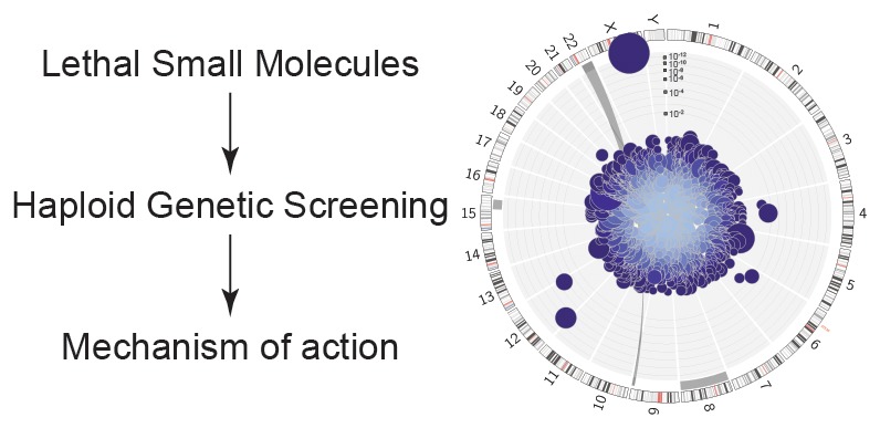
          Little is known about the regulation of nonapoptotic cell death. Using massive insertional mutagenesis of haploid KBM7 cells we identified nine genes involved in small-molecule-induced nonapoptotic cell death, including mediators of fatty acid metabolism ( ACSL4) and lipid remodeling ( LPCAT3) in ferroptosis. One novel compound, CIL56, triggered cell death dependent upon the rate-limiting de novo lipid synthetic enzyme ACC1. These results provide insight into the genetic regulation of cell death and highlight the central role of lipid metabolism in nonapoptotic cell death.

          Related collections

          Most cited references16

          • Record: found
          • Abstract: found
          • Article: not found

          RAS-RAF-MEK-dependent oxidative cell death involving voltage-dependent anion channels.

          Therapeutics that discriminate between the genetic makeup of normal cells and tumour cells are valuable for treating and understanding cancer. Small molecules with oncogene-selective lethality may reveal novel functions of oncoproteins and enable the creation of more selective drugs. Here we describe the mechanism of action of the selective anti-tumour agent erastin, involving the RAS-RAF-MEK signalling pathway functioning in cell proliferation, differentiation and survival. Erastin exhibits greater lethality in human tumour cells harbouring mutations in the oncogenes HRAS, KRAS or BRAF. Using affinity purification and mass spectrometry, we discovered that erastin acts through mitochondrial voltage-dependent anion channels (VDACs)--a novel target for anti-cancer drugs. We show that erastin treatment of cells harbouring oncogenic RAS causes the appearance of oxidative species and subsequent death through an oxidative, non-apoptotic mechanism. RNA-interference-mediated knockdown of VDAC2 or VDAC3 caused resistance to erastin, implicating these two VDAC isoforms in the mechanism of action of erastin. Moreover, using purified mitochondria expressing a single VDAC isoform, we found that erastin alters the permeability of the outer mitochondrial membrane. Finally, using a radiolabelled analogue and a filter-binding assay, we show that erastin binds directly to VDAC2. These results demonstrate that ligands to VDAC proteins can induce non-apoptotic cell death selectively in some tumour cells harbouring activating mutations in the RAS-RAF-MEK pathway.
            Bookmark
            • Record: found
            • Abstract: found
            • Article: not found

            Synchronized renal tubular cell death involves ferroptosis.

            Receptor-interacting protein kinase 3 (RIPK3)-mediated necroptosis is thought to be the pathophysiologically predominant pathway that leads to regulated necrosis of parenchymal cells in ischemia-reperfusion injury (IRI), and loss of either Fas-associated protein with death domain (FADD) or caspase-8 is known to sensitize tissues to undergo spontaneous necroptosis. Here, we demonstrate that renal tubules do not undergo sensitization to necroptosis upon genetic ablation of either FADD or caspase-8 and that the RIPK1 inhibitor necrostatin-1 (Nec-1) does not protect freshly isolated tubules from hypoxic injury. In contrast, iron-dependent ferroptosis directly causes synchronized necrosis of renal tubules, as demonstrated by intravital microscopy in models of IRI and oxalate crystal-induced acute kidney injury. To suppress ferroptosis in vivo, we generated a novel third-generation ferrostatin (termed 16-86), which we demonstrate to be more stable, to metabolism and plasma, and more potent, compared with the first-in-class compound ferrostatin-1 (Fer-1). Even in conditions with extraordinarily severe IRI, 16-86 exerts strong protection to an extent which has not previously allowed survival in any murine setting. In addition, 16-86 further potentiates the strong protective effect on IRI mediated by combination therapy with necrostatins and compounds that inhibit mitochondrial permeability transition. Renal tubules thus represent a tissue that is not sensitized to necroptosis by loss of FADD or caspase-8. Finally, ferroptosis mediates postischemic and toxic renal necrosis, which may be therapeutically targeted by ferrostatins and by combination therapy.
              Bookmark
              • Record: found
              • Abstract: found
              • Article: not found

              Ferrostatins Inhibit Oxidative Lipid Damage and Cell Death in Diverse Disease Models

              Ferrostatin-1 (Fer-1) inhibits ferroptosis, a form of regulated, oxidative, nonapoptotic cell death. We found that Fer-1 inhibited cell death in cellular models of Huntington’s disease (HD), periventricular leukomalacia (PVL), and kidney dysfunction; Fer-1 inhibited lipid peroxidation, but not mitochondrial reactive oxygen species formation or lysosomal membrane permeability. We developed a mechanistic model to explain the activity of Fer-1, which guided the development of ferrostatins with improved properties. These studies suggest numerous therapeutic uses for ferrostatins, and that lipid peroxidation mediates diverse disease phenotypes.
                Bookmark

                Author and article information

                Journal
                ACS Chem Biol
                ACS Chem. Biol
                cb
                acbcct
                ACS Chemical Biology
                American Chemical Society
                1554-8929
                1554-8937
                12 May 2016
                12 May 2015
                17 July 2015
                : 10
                : 7
                : 1604-1609
                Affiliations
                [1] Department of Biological Sciences, Department of Chemistry, and §Howard Hughes Medical Institute, Columbia University , 550 West 120th Street, Northwest Corner Building, MC 4846, New York, New York 10027, United States
                []CeMM Research Center for Molecular Medicine of the Austrian Academy of Sciences , Lazarettgasse 14, 1090 Vienna, Austria
                []Department of Biology, Stanford University , 337 Campus Drive, Stanford, California 94305, United States
                Author notes
                Article
                10.1021/acschembio.5b00245
                4509420
                25965523
                b7bbb6cf-2e65-4292-b0ab-547bd97d9902
                Copyright © 2015 American Chemical Society

                This is an open access article published under an ACS AuthorChoice License, which permits copying and redistribution of the article or any adaptations for non-commercial purposes.

                History
                : 06 April 2015
                : 12 May 2015
                Categories
                Letters
                Custom metadata
                cb5b00245
                cb-2015-002457

                Biochemistry
                Biochemistry

                Comments

                Comment on this article