55
views
0
recommends
+1 Recommend
0 collections
    0
    shares
      • Record: found
      • Abstract: found
      • Article: found
      Is Open Access

      The Regulation of Steroid Action by Sulfation and Desulfation

      review-article

      Read this article at

      Bookmark
          There is no author summary for this article yet. Authors can add summaries to their articles on ScienceOpen to make them more accessible to a non-specialist audience.

          Abstract

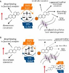
          Steroid sulfation and desulfation are fundamental pathways vital for a functional vertebrate endocrine system. After biosynthesis, hydrophobic steroids are sulfated to expedite circulatory transit. Target cells express transmembrane organic anion-transporting polypeptides that facilitate cellular uptake of sulfated steroids. Once intracellular, sulfatases hydrolyze these steroid sulfate esters to their unconjugated, and usually active, forms. Because most steroids can be sulfated, including cholesterol, pregnenolone, dehydroepiandrosterone, and estrone, understanding the function, tissue distribution, and regulation of sulfation and desulfation processes provides significant insights into normal endocrine function. Not surprisingly, dysregulation of these pathways is associated with numerous pathologies, including steroid-dependent cancers, polycystic ovary syndrome, and X-linked ichthyosis. Here we provide a comprehensive examination of our current knowledge of endocrine-related sulfation and desulfation pathways. We describe the interplay between sulfatases and sulfotransferases, showing how their expression and regulation influences steroid action. Furthermore, we address the role that organic anion-transporting polypeptides play in regulating intracellular steroid concentrations and how their expression patterns influence many pathologies, especially cancer. Finally, the recent advances in pharmacologically targeting steroidogenic pathways will be examined.

          Related collections

          Most cited references390

          • Record: found
          • Abstract: found
          • Article: not found

          The complex role of estrogens in inflammation.

          There is still an unresolved paradox with respect to the immunomodulating role of estrogens. On one side, we recognize inhibition of bone resorption and suppression of inflammation in several animal models of chronic inflammatory diseases. On the other hand, we realize the immunosupportive role of estrogens in trauma/sepsis and the proinflammatory effects in some chronic autoimmune diseases in humans. This review examines possible causes for this paradox. This review delineates how the effects of estrogens are dependent on criteria such as: 1) the immune stimulus (foreign antigens or autoantigens) and subsequent antigen-specific immune responses (e.g., T cell inhibited by estrogens vs. activation of B cell); 2) the cell types involved during different phases of the disease; 3) the target organ with its specific microenvironment; 4) timing of 17beta-estradiol administration in relation to the disease course (and the reproductive status of a woman); 5) the concentration of estrogens; 6) the variability in expression of estrogen receptor alpha and beta depending on the microenvironment and the cell type; and 7) intracellular metabolism of estrogens leading to important biologically active metabolites with quite different anti- and proinflammatory function. Also mentioned are systemic supersystems such as the hypothalamic-pituitary-adrenal axis, the sensory nervous system, and the sympathetic nervous system and how they are influenced by estrogens. This review reinforces the concept that estrogens have antiinflammatory but also proinflammatory roles depending on above-mentioned criteria. It also explains that a uniform concept as to the action of estrogens cannot be found for all inflammatory diseases due to the enormous variable responses of immune and repair systems.
            Bookmark
            • Record: found
            • Abstract: found
            • Article: not found

            Endogenous sex hormones and prostate cancer: a collaborative analysis of 18 prospective studies.

            Sex hormones in serum have been hypothesized to influence the risk of prostate cancer. We performed a collaborative analysis of the existing worldwide epidemiologic data to examine these associations in a uniform manner and to provide more precise estimates of risks. Data on serum concentrations of sex hormones from 18 prospective studies that included 3886 men with incident prostate cancer and 6438 control subjects were pooled by the Endogenous Hormones and Prostate Cancer Collaborative Group. Relative risks (RRs) of prostate cancer by fifths of serum hormone concentration were estimated by use of conditional logistic regression with stratification by study, age at recruitment, and year of recruitment. All statistical tests were two-sided. No associations were found between the risk of prostate cancer and serum concentrations of testosterone, calculated free testosterone, dihydrotestosterone, dehydroepiandrosterone sulfate, androstenedione, androstanediol glucuronide, estradiol, or calculated free estradiol. The serum concentration of sex hormone-binding globulin was modestly inversely associated with prostate cancer risk (RR in the highest vs lowest fifth = 0.86, 95% confidence interval = 0.75 to 0.98; P(trend) = .01). There was no statistical evidence of heterogeneity among studies, and adjustment for potential confounders made little difference to the risk estimates. In this collaborative analysis of the worldwide data on endogenous hormones and prostate cancer risk, serum concentrations of sex hormones were not associated with the risk of prostate cancer.
              Bookmark
              • Record: found
              • Abstract: found
              • Article: not found

              Estrogens protect against high-fat diet-induced insulin resistance and glucose intolerance in mice.

              Although corroborating data indicate that estrogens influence glucose metabolism through the activation of the estrogen receptor alpha (ERalpha), it has not been established whether this pathway could represent an effective therapeutic target to fight against metabolic disturbances induced by a high-fat diet (HFD). To this end, we first evaluated the influence of chronic 17beta-estradiol (E2) administration in wild-type ovariectomized mice submitted to either a normal chow diet or a HFD. Whereas only a modest effect was observed in normal chow diet-fed mice, E2 administration exerted a protective effect against HFD-induced glucose intolerance, and this beneficial action was abolished in ERalpha-deficient mice. Furthermore, E2 treatment reduced HFD-induced insulin resistance by 50% during hyperinsulinemic euglycemic clamp studies and improved insulin signaling (Akt phosphorylation) in insulin-stimulated skeletal muscles. Unexpectedly, we found that E2 treatment enhanced cytokine (IL-6, TNF-alpha) and plasminogen activator inhibitor-1 mRNA expression induced by HFD in the liver and visceral adipose tissue. Interestingly, although the proinflammatory effect of E2 was abolished in visceral adipose tissue from chimeric mice grafted with bone marrow cells from ERalpha-deficient mice, the beneficial effect of the hormone on glucose tolerance was not altered, suggesting that the metabolic and inflammatory effects of estrogens can be dissociated. Eventually comparison of sham-operated with ovariectomized HFD-fed mice demonstrated that endogenous estrogens levels are sufficient to exert a full protective effect against insulin resistance and glucose intolerance. In conclusion, the regulation of the ERalpha pathway could represent an effective strategy to reduce the impact of high-fat diet-induced type 2 diabetes.
                Bookmark

                Author and article information

                Journal
                Endocr Rev
                Endocr. Rev
                edrv
                endre
                edrv
                edrv
                Endocrine Reviews
                Endocrine Society (Washington, DC )
                0163-769X
                1945-7189
                October 2015
                27 July 2015
                27 July 2015
                : 36
                : 5
                : 526-563
                Affiliations
                Centre for Endocrinology, Diabetes, and Metabolism, Institute of Metabolism and Systems Research, University of Birmingham, Birmingham B15 2TT, United Kingdom
                Author notes
                Address all correspondence and requests for reprints to: Paul Alexander Foster, PhD, Centre for Endocrinology, Diabetes, and Metabolism, Institute of Metabolism and Systems Research, University of Birmingham, Birmingham B15 2TT, United Kingdom. E-mail: p.a.foster@ 123456bham.ac.uk .
                Article
                ER-15-1036
                10.1210/er.2015-1036
                4591525
                26213785
                b72b1b2a-b24c-468c-8936-e40145d3ff51

                This article has been published under the terms of the Creative Commons Attribution License (CC-BY; http://creativecommons.org/licenses/by/4.0/), which permits unrestricted use, distribution, and reproduction in any medium, provided the original author and source are credited. Copyright for this article is retained by the author(s).

                History
                : 30 March 2015
                : 21 July 2015
                Categories
                Reviews

                Comments

                Comment on this article