0
views
0
recommends
+1 Recommend
0 collections
    0
    shares
      • Record: found
      • Abstract: found
      • Article: found
      Is Open Access

      Reduced Dehydroepiandrosterone-Sulfate Levels in the Mid-Luteal Subphase of the Menstrual Cycle: Implications to Women’s Health Research

      , , , , ,
      Metabolites
      MDPI AG

      Read this article at

      Bookmark
          There is no author summary for this article yet. Authors can add summaries to their articles on ScienceOpen to make them more accessible to a non-specialist audience.

          Abstract

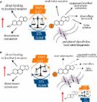
          The regulation of DHEA-sulfate by steroid sulfotransferase (SULT) and steryl-sulfatase (STS) enzymes is a vital process for the downstream formation of many steroid hormones. DHEA-sulfate is the most abundant steroid hormone in the human body; thus, DHEA-sulfate and its hydrolyzed form, DHEA, continue to be evaluated in numerous studies, given their importance to human health. Yet, a basic question of relevance to the reproductive-age female population—whether the two steroid hormones vary across the menstrual cycle—has not been addressed. We applied a validated, multi-step protocol, involving realignment and imputation of study data to early follicular, mid-late follicular, periovulatory, and early, mid-, and late luteal subphases of the menstrual cycle, and analyzed DHEA-sulfate and DHEA serum concentrations using ultraperformance liquid chromatography tandem mass spectrometry. DHEA-sulfate levels started to decrease in the early luteal, significantly dropped in the mid-luteal, and returned to basal levels by the late luteal subphase. DHEA, however, did not vary across the menstrual cycle. The present study deep-mapped trajectories of DHEA and DHEA-sulfate across the entire menstrual cycle, demonstrating a significant decrease in DHEA-sulfate in the mid-luteal subphase. These findings are relevant to the active area of research examining associations between DHEA-sulfate levels and various disease states.

          Related collections

          Most cited references52

          • Record: found
          • Abstract: found
          • Article: not found

          Neurobiological and neuropsychiatric effects of dehydroepiandrosterone (DHEA) and DHEA sulfate (DHEAS).

          DHEA and DHEAS are steroids synthesized in human adrenals, but their function is unclear. In addition to adrenal synthesis, evidence also indicates that DHEA and DHEAS are synthesized in the brain, further suggesting a role of these hormones in brain function and development. Despite intensifying research into the biology of DHEA and DHEAS, many questions concerning their mechanisms of action and their potential involvement in neuropsychiatric illnesses remain unanswered. We review and distill the preclinical and clinical data on DHEA and DHEAS, focusing on (i) biological actions and putative mechanisms of action, (ii) differences in endogenous circulating concentrations in normal subjects and patients with neuropsychiatric diseases, and (iii) the therapeutic potential of DHEA in treating these conditions. Biological actions of DHEA and DHEAS include neuroprotection, neurite growth, and antagonistic effects on oxidants and glucocorticoids. Accumulating data suggest abnormal DHEA and/or DHEAS concentrations in several neuropsychiatric conditions. The evidence that DHEA and DHEAS may be fruitful targets for pharmacotherapy in some conditions is reviewed.
            Bookmark
            • Record: found
            • Abstract: found
            • Article: found
            Is Open Access

            The Regulation of Steroid Action by Sulfation and Desulfation

            Steroid sulfation and desulfation are fundamental pathways vital for a functional vertebrate endocrine system. After biosynthesis, hydrophobic steroids are sulfated to expedite circulatory transit. Target cells express transmembrane organic anion-transporting polypeptides that facilitate cellular uptake of sulfated steroids. Once intracellular, sulfatases hydrolyze these steroid sulfate esters to their unconjugated, and usually active, forms. Because most steroids can be sulfated, including cholesterol, pregnenolone, dehydroepiandrosterone, and estrone, understanding the function, tissue distribution, and regulation of sulfation and desulfation processes provides significant insights into normal endocrine function. Not surprisingly, dysregulation of these pathways is associated with numerous pathologies, including steroid-dependent cancers, polycystic ovary syndrome, and X-linked ichthyosis. Here we provide a comprehensive examination of our current knowledge of endocrine-related sulfation and desulfation pathways. We describe the interplay between sulfatases and sulfotransferases, showing how their expression and regulation influences steroid action. Furthermore, we address the role that organic anion-transporting polypeptides play in regulating intracellular steroid concentrations and how their expression patterns influence many pathologies, especially cancer. Finally, the recent advances in pharmacologically targeting steroidogenic pathways will be examined.
              Bookmark
              • Record: found
              • Abstract: found
              • Article: not found

              Peripheral BDNF produces antidepressant-like effects in cellular and behavioral models.

              Recent clinical studies demonstrate that serum levels of brain-derived neurotrophic factor (BDNF) are significantly decreased in patients with major depressive disorder (MDD) and that antidepressant treatments reverse this effect, indicating that serum BDNF is a biomarker of MDD. These findings raise the possibility that serum BDNF may also have effects on neuronal activity and behavior, but the functional significance of altered serum BDNF is unknown. To address this issue, we determined the influence of peripheral BDNF administration on depression- and anxiety-like behavior, including the forced swim test (FST), chronic unpredictable stress (CUS)/anhedonia, novelty-induced hypophagia (NIH) test, and elevated-plus maze (EPM). Furthermore, we examined adult hippocampal neurogenesis as well as hippocampal and striatal expression of BDNF, extracellular signal-regulated kinase (ERK) and cAMP response element-binding protein (CREB), in order to determine whether peripherally administered BDNF produces antidepressant-like cellular responses in the brain. Peripheral BDNF administration increased mobility in the FST, attenuated the effects of CUS on sucrose consumption, decreased latency in the NIH test, and increased time spent in the open arms of an EPM. Moreover, adult hippocampal neurogenesis was increased after chronic, peripheral BDNF administration. We also found that BDNF levels as well as expression of pCREB and pERK were elevated in the hippocampus of adult mice receiving peripheral BDNF. Taken together, these results indicate that peripheral/serum BDNF may not only represent a biomarker of MDD, but also have functional consequences on molecular signaling substrates, neurogenesis, and behavior.
                Bookmark

                Author and article information

                Contributors
                Journal
                METALU
                Metabolites
                Metabolites
                MDPI AG
                2218-1989
                October 2022
                October 04 2022
                : 12
                : 10
                : 941
                Article
                10.3390/metabo12100941
                36295844
                3a3ed221-5cc3-470d-b5a1-98b25c4f59fd
                © 2022

                https://creativecommons.org/licenses/by/4.0/

                History

                Comments

                Comment on this article