5
views
0
recommends
+1 Recommend
0 collections
    0
    shares
      • Record: found
      • Abstract: found
      • Article: found
      Is Open Access

      LncRNA-GAS5 promotes spinal cord repair and the inhibition of neuronal apoptosis via the transplantation of 3D printed scaffold loaded with induced pluripotent stem cell-derived neural stem cells

      research-article

      Read this article at

      Bookmark
          There is no author summary for this article yet. Authors can add summaries to their articles on ScienceOpen to make them more accessible to a non-specialist audience.

          Abstract

          Background

          Stem cell transplantation has been increasingly used for spinal cord repair, and some achievements have been made. However, limited stem cell sources as well as immune rejection and ethical issues have restricted its wide application. Therefore, to achieve further breakthroughs regarding the application of stem cell transplantation to treat spinal cord injury (SCI), it is important to develop a stem cell line that can effectively avoid immune rejection and ethical issues.

          Methods

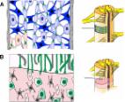
          Urine cells (UCs) were induced to differentiate into induced pluripotent stem cells (iPSCs), which then further differentiated into neural stem cells (NSCs). Relevant tests were performed, and three-dimensional (3D) printed scaffolds were prepared. Thirty C57BL/6 mice were divided into 5 groups based on a random number table: a sham group, an SCI group, an SCI + control group, an SCI + siNC group, and an SCI + siGAS5 group (n=6). The latter 4 groups replicated SCI models. Mice in the SCI + control group were transplanted with 3D scaffolds loaded with iPSC-derived NSCs (iPSd-NSCs). Mice in the SCI + siNC group and the SCI + siGAS5 group were transplanted with scaffolds loaded with iPSd-NSCs-siNC and 3D scaffolds loaded with iPSd-NSCs-siGAS5, respectively. Mice in the other groups were injected with the same amount of normal saline. Hematoxylin-eosin staining was used to observe the histopathology of the injured spinal cord, the Basso-Mouse Scale was used to assess the motor function of the hind limbs of the mice, and Western blot was used to detect the expression of apoptosis-related proteins after SCI.

          Results

          iPSd-NSCs were successfully induced and differentiated, and 3D printed heparin sulfate-collagen scaffolds were prepared, inside which a 3D loose porous structure was shown by electron microscopy. Morphological observations showed that iPSd-NSC transplantation improved SCI in mice, while GAS5 silencing inhibited the reparative effect of iPSd-NSC transplantation on SCI in mice. Western blot results indicated that iPSd-NSC transplantation significantly increased the expression level of B cell lymphoma/leukemia-2 (Bcl-2) (P<0.01) but decreased the expression levels of Bcl-2 associated X protein, cytochrome C, and cleaved caspase-3 (P<0.001).

          Conclusions

          The overexpression of lncRNA-GAS5 can promote spinal cord repair and inhibit neural apoptosis via the transplantation of 3D printed scaffolds loaded with iPSd-NSCs.

          Related collections

          Most cited references16

          • Record: found
          • Abstract: found
          • Article: not found

          Generation of induced pluripotent stem cells from urine.

          Forced expression of selected transcription factors can transform somatic cells into embryonic stem cell (ESC)-like cells, termed induced pluripotent stem cells (iPSCs). There is no consensus regarding the preferred tissue from which to harvest donor cells for reprogramming into iPSCs, and some donor cell types may be more prone than others to accumulation of epigenetic imprints and somatic cell mutations. Here, we present a simple, reproducible, noninvasive method for generating human iPSCs from renal tubular cells present in urine. This procedure eliminates many problems associated with other protocols, and the resulting iPSCs display an excellent ability to differentiate. These data suggest that urine may be a preferred source for generating iPSCs. Copyright © 2011 by the American Society of Nephrology
            Bookmark
            • Record: found
            • Abstract: found
            • Article: not found

            iPS cells reprogrammed from human mesenchymal-like stem/progenitor cells of dental tissue origin.

            Generation of induced pluripotent stem (iPS) cells holds a great promise for regenerative medicine and other aspects of clinical applications. Many types of cells have been successfully reprogrammed into iPS cells in the mouse system; however, reprogramming human cells have been more difficult. To date, human dermal fibroblasts are the most accessible and feasible cell source for iPS generation. Dental tissues derived from ectomesenchyme harbor mesenchymal-like stem/progenitor cells and some of the tissues have been treated as biomedical wastes, for example, exfoliated primary teeth and extracted third molars. We asked whether stem/progenitor cells from discarded dental tissues can be reprogrammed into iPS cells. The 4 factors Lin28/Nanog/Oct4/Sox2 or c-Myc/Klf4/Oct4/Sox2 carried by viral vectors were used to reprogram 3 different dental stem/progenitor cells: stem cells from exfoliated deciduous teeth (SHED), stem cells from apical papilla (SCAP), and dental pulp stem cells (DPSCs). We showed that all 3 can be reprogrammed into iPS cells and appeared to be at a higher rate than fibroblasts. They exhibited a morphology indistinguishable from human embryonic stem (hES) cells in cultures and expressed hES cell markers SSEA-4, TRA-1-60, TRA-1-80, TRA-2-49, Nanog, Oct4, and Sox2. They formed embryoid bodies in vitro and teratomas in vivo containing tissues of all 3 germ layers. We conclude that cells of ectomesenchymal origin serve as an excellent alternative source for generating iPS cells.
              Bookmark
              • Record: found
              • Abstract: found
              • Article: found
              Is Open Access

              Repair of injured spinal cord using biomaterial scaffolds and stem cells

              The loss of neurons and degeneration of axons after spinal cord injury result in the loss of sensory and motor functions. A bridging biomaterial construct that allows the axons to grow through has been investigated for the repair of injured spinal cord. Due to the hostility of the microenvironment in the lesion, multiple conditions need to be fulfilled to achieve improved functional recovery. A scaffold has been applied to bridge the gap of the lesion as contact guidance for axonal growth and to act as a vehicle to deliver stem cells in order to modify the microenvironment. Stem cells may improve functional recovery of the injured spinal cord by providing trophic support or directly replacing neurons and their support cells. Neural stem cells and mesenchymal stem cells have been seeded into biomaterial scaffolds and investigated for spinal cord regeneration. Both natural and synthetic biomaterials have increased stem cell survival in vivo by providing the cells with a controlled microenvironment in which cell growth and differentiation are facilitated. This optimal multi‒disciplinary approach of combining biomaterials, stem cells, and biomolecules offers a promising treatment for the injured spinal cord.
                Bookmark

                Author and article information

                Journal
                Ann Transl Med
                Ann Transl Med
                ATM
                Annals of Translational Medicine
                AME Publishing Company
                2305-5839
                2305-5847
                June 2021
                June 2021
                : 9
                : 11
                : 931
                Affiliations
                [1 ]deptDepartment of Orthopedics , Hangzhou TCM Hospital Affiliated to Zhejiang Chinese Medical University , Hangzhou, China;
                [2 ]deptDepartment of Orthopedics , Jiangnan Hospital Affiliated to Zhejiang Chinese Medical University , Hangzhou, China;
                [3 ]deptOrthopaedic Institute , Zhejiang Chinese Medical University , Hangzhou, China
                Author notes

                Contributions: (I) Conception and design: R Shao; (II) Administrative support: J Yue, H Zhu, H Pan, H Zhou, R Quan; (III) Provision of study materials or patients: L Zhang, H Yang, Z Zhang; (IV) Collection and assembly of data: Y Chen, W Gao; (V) Data analysis and interpretation: C Li; (VI) Manuscript writing: All authors; (VII) Final approval of manuscript: All authors.

                [#]

                These authors contributed equally to this work.

                Correspondence to: Rongxue Shao. Department of Orthopedics, Hangzhou TCM Hospital Affiliated to Zhejiang Chinese Medical University, No. 453, Tiyuchang Road, Hangzhou 310007, China. Email: shaorongxue@ 123456163.com ; Changming Li. Department of Orthopedics, Jiangnan Hospital Affiliated to Zhejiang Chinese Medical University, No. 152, Yucai Road, Hangzhou 311200, China. Email: lcmmail@ 123456126.com .
                Article
                atm-09-11-931
                10.21037/atm-21-2570
                8263850
                34350246
                b6c1d0c7-22c7-4bfb-869d-bd37f87b56fa
                2021 Annals of Translational Medicine. All rights reserved.

                Open Access Statement: This is an Open Access article distributed in accordance with the Creative Commons Attribution-NonCommercial-NoDerivs 4.0 International License (CC BY-NC-ND 4.0), which permits the non-commercial replication and distribution of the article with the strict proviso that no changes or edits are made and the original work is properly cited (including links to both the formal publication through the relevant DOI and the license). See: https://creativecommons.org/licenses/by-nc-nd/4.0.

                History
                : 08 April 2021
                : 03 June 2021
                Categories
                Original Article

                long non-coding rna-gas5,3d printed scaffold,ipsc-derived nscs,spinal cord injury (sci),neuronal apoptosis

                Comments

                Comment on this article