0
views
0
recommends
+1 Recommend
0 collections
    0
    shares
      • Record: found
      • Abstract: found
      • Article: found
      Is Open Access

      Accuracy of end-on fluoroscopy in predicting implant position in relation to the vertebral canal in dogs

      research-article

      Read this article at

      Bookmark
          There is no author summary for this article yet. Authors can add summaries to their articles on ScienceOpen to make them more accessible to a non-specialist audience.

          Abstract

          Objective

          To evaluate the accuracy of end-on fluoroscopy in predicting implant position in relation to the vertebral canal in the canine thoracolumbar vertebral column.

          Study design

          In vitro imaging and anatomic study.

          Animals

          Canine cadaveric thoracolumbar vertebral columns ( n = 5).

          Methods

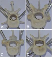
          Smooth Steinmann pins were inserted bicortically into the thoracolumbar vertebral columns between T10 and L7 using recommended insertion angles. Penetration of the spinal canal was not strictly avoided. After pin placement, end-on fluoroscopy images were obtained of each pin. Pin position was subsequently assessed by four evaluators and determined to either being out of the vertebral canal or in, with the latter being additionally divided into partially or completely penetrating the canal. To assess potential differences in modalities, fluoroscopy images were gray-scale inverted and evaluated again later by the same four individuals. Correct identification of pin position in relationship to the vertebral canal was assessed for both fluoroscopy images. Anatomic preparation of the spines was used for verification of pin position in relation to the spinal canal. Some data from this study were compared with historical data on accuracy using orthogonal radiography and computed tomography (CT).

          Results

          Overall sensitivity and specificity of F to detect vertebral canal penetration was 98.8 % (95% confidence interval (CI), 96.0–99.6) and 98.0% (95% CI, 77.0–99.9), respectively. For Fi, sensitivity and specificity were 97.0% (95% CI, 91.5–99.0) and 98.5% (95% CI, 81.5–99.9) respectively. F exceeded Fi for the sensitivity of detecting pin penetration into the vertebral canal ( p = 0.039) but specificities were not different ( p = 0.585). When comparing to historical data, the overall accuracy of end-on fluoroscopy (F) and inverted fluoroscopy (Fi) was statistical better than conventional radiographic assessment ( p < 0.001).

          Conclusion

          End-on fluoroscopy is a highly accurate method for the assessment of pin position in relationship to the thoracolumbar spinal canal in cadaveric dogs.

          Clinical significance

          End-on fluoroscopy, with or without inversion, is accurate in identifying vertebral canal violation by bicortically placed Steinmann pins. When CT is not available, end-on fluoroscopy might be a valuable imaging modality to determine pin position in the canine vertebral column.

          Related collections

          Most cited references32

          • Record: found
          • Abstract: found
          • Article: not found

          Safety and accuracy of robot-assisted versus fluoroscopy-assisted pedicle screw insertion in thoracolumbar spinal surgery: a prospective randomized controlled trial

          The object of this study was to compare the safety and accuracy of pedicle screw placement using the TiRobot system versus conventional fluoroscopy in thoracolumbar spinal surgery. Patients with degenerative or traumatic thoracolumbar spinal disorders requiring spinal instrumentation were randomly assigned to either the TiRobot-assisted group (RG) or the freehand fluoroscopy-assisted group (FG) at a 1:1 ratio. The primary outcome measure was the accuracy of screw placement according to the Gertzbein-Robbins scale; grades A and B (pedicle breach < 2 mm) were considered clinically acceptable. In the RG, discrepancies between the planned and actual screw placements were measured by merging postoperative CT images with the trajectory planning images. Secondary outcome parameters included proximal facet joint violation, duration of surgery, intraoperative blood loss, conversion to freehand approach in the RG, postoperative hospital stay, and radiation exposure. A total of 1116 pedicle screws were implanted in 234 patients (119 in the FG, and 115 in the RG). In the RG, 95.3% of the screws were perfectly positioned (grade A); the remaining screws were graded B (3.4%), C (0.9%), and D (0.4%). In the FG, 86.1% screws were perfectly positioned (grade A); the remaining screws were graded B (7.4%), C (4.6%), D (1.4%), and E (0.5%). The proportion of clinically acceptable screws was significantly greater in the RG than in the FG (p < 0.01). In the RG, the mean deviation was 1.5 ± 0.8 mm for each screw. The most common direction of screw deviation was lateral in the RG and medial in the FG. Two misplaced screws in the FG required revision surgery, whereas no revision was required in the RG. None of the screws in the RG violated the proximal facet joint, whereas 12 screws (2.1%) in the FG violated the proximal facet joint (p < 0.01). The RG had significantly less blood loss (186.0 ± 255.3 ml) than the FG (217.0 ± 174.3 ml; p < 0.05). There were no significant differences between the two groups in terms of surgical time and postoperative hospital stay. The mean cumulative radiation time was 81.5 ± 38.6 seconds in the RG and 71.5 ± 44.2 seconds in the FG (p = 0.07). Surgeon radiation exposure was significantly less in the RG (21.7 ± 11.5 μSv) than in the FG (70.5 ± 42.0 μSv; p < 0.01). TiRobot-guided pedicle screw placement is safe and useful in thoracolumbar spinal surgery. Clinical trial registration no.: NCT02890043 (clinicaltrials.gov)
            Bookmark
            • Record: found
            • Abstract: found
            • Article: not found

            A prospective study of postoperative surgical site infections in dogs and cats.

            To assess postoperative surgical site infection (SSI) rate and to identify associated predictive factors. Prospective clinical study. Dogs and cats that had surgery (1010 interventions) during 58 weeks from April 1999 to June 2000. Data sheets were completed by clinicians. Patients were controlled for clinical evidence of SSI at suture removal. Two definitions of SSI ("infection" and "infection/inflammation") were developed specifically for this study and used for statistical analysis. Logistic regression models were built in order to identify significant predictive factors for SSI. Wounds with "infection/inflammation" occurred in 5.8% and "infected" wounds in 3% of patients. The outcome "infection" was associated with 3 major risk factors (duration of surgery, increasing number of persons in the operating room, dirty surgical site) and 1 protective factor (antimicrobial prophylaxis). The outcome "infection/inflammation" was associated with 6 significant factors (duration of anesthesia, duration of postoperative intensive care unit stay, wound drainage, increasing patient weight, dirty surgical site, and antimicrobial prophylaxis). SSI frequency in companion animals is comparable with the frequency observed in human surgical patients. Several significant predictive factors for SSI in small animals surgery were identified. Baseline information for SSI surveillance in our hospital and for comparison with other studies was defined. The factors identified may help to predict infections in surgical patients and to take adequate preventive measures for patients at risk.
              Bookmark
              • Record: found
              • Abstract: found
              • Article: found
              Is Open Access

              Accuracy of pedicle screw placement comparing robot-assisted technology and the free-hand with fluoroscopy-guided method in spine surgery

              Supplemental Digital Content is available in the text
                Bookmark

                Author and article information

                Contributors
                Journal
                Front Vet Sci
                Front Vet Sci
                Front. Vet. Sci.
                Frontiers in Veterinary Science
                Frontiers Media S.A.
                2297-1769
                20 October 2022
                2022
                : 9
                : 982560
                Affiliations
                [1] 1Department of Clinical Veterinary Medicine, Vetsuisse Faculty, University of Bern , Bern, Switzerland
                [2] 2Department of Production Animal Studies, University of Pretoria, Onderstepoort , Pretoria, South Africa
                Author notes

                Edited by: Andrea Tipold, University of Veterinary Medicine Hannover, Germany

                Reviewed by: Karolin Campbell, Radiovet, Switzerland; Andrea Meyer-Lindenberg, Ludwig Maximilian University of Munich, Germany

                *Correspondence: Bianca F. Hettlich bhettlich@ 123456gmail.com

                This article was submitted to Veterinary Neurology and Neurosurgery, a section of the journal Frontiers in Veterinary Science

                Article
                10.3389/fvets.2022.982560
                9630941
                36337187
                b5b60d2d-09e3-4154-92bd-517082659d11
                Copyright © 2022 Goffart, Precht, Fosgate, Maiolini and Hettlich.

                This is an open-access article distributed under the terms of the Creative Commons Attribution License (CC BY). The use, distribution or reproduction in other forums is permitted, provided the original author(s) and the copyright owner(s) are credited and that the original publication in this journal is cited, in accordance with accepted academic practice. No use, distribution or reproduction is permitted which does not comply with these terms.

                History
                : 30 June 2022
                : 29 September 2022
                Page count
                Figures: 4, Tables: 3, Equations: 0, References: 32, Pages: 9, Words: 6029
                Categories
                Veterinary Science
                Original Research

                vertebral column,canine,bicortical pins,accuracy,fluoroscopy,end-on,gray-scale inversion

                Comments

                Comment on this article