14
views
0
recommends
+1 Recommend
0 collections
    0
    shares
      • Record: found
      • Abstract: found
      • Article: found
      Is Open Access

      Central vein sign differentiates Multiple Sclerosis from central nervous system inflammatory vasculopathies

      research-article

      Read this article at

      Bookmark
          There is no author summary for this article yet. Authors can add summaries to their articles on ScienceOpen to make them more accessible to a non-specialist audience.

          Abstract

          Objectives

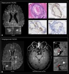
          In multiple sclerosis (MS), magnetic resonance imaging (MRI) is a sensitive tool for detecting white matter lesions, but its diagnostic specificity is still suboptimal; ambiguous cases are frequent in clinical practice. Detection of perivenular lesions in the brain (the “central vein sign”) improves the pathological specificity of MS diagnosis, but comprehensive evaluation of this MRI biomarker in MS‐mimicking inflammatory and/or autoimmune diseases, such as central nervous system (CNS) inflammatory vasculopathies, is lacking. In a multicenter study, we assessed the frequency of perivenular lesions in MS versus systemic autoimmune diseases with CNS involvement and primary angiitis of the CNS (PACNS).

          Methods

          In 31 patients with inflammatory CNS vasculopathies and 52 with relapsing–remitting MS, 3‐dimensional T2*‐weighted and T2–fluid‐attenuated inversion recovery images were obtained during a single MRI acquisition after gadolinium injection. For each lesion, the central vein sign was evaluated according to consensus guidelines. For each patient, lesion count, volume, and brain location, as well as fulfillment of dissemination in space MRI criteria, were assessed.

          Results

          MS showed higher frequency of perivenular lesions (median = 88%) than did inflammatory CNS vasculopathies (14%), without overlap between groups or differences between 3T and 1.5T MRI. Among inflammatory vasculopathies, Behçet disease showed the highest median frequency of perivenular lesions (34%), followed by PACNS (14%), antiphospholipid syndromes (12%), Sjögren syndrome (11%), and systemic lupus erythematosus (0%). When a threshold of 50% perivenular lesions was applied, central vein sign discriminated MS from inflammatory vasculopathies with a diagnostic accuracy of 100%.

          Interpretation

          The central vein sign differentiates inflammatory CNS vasculopathies from MS at standard clinical magnetic field strengths. Ann Neurol 2018;83:283–294

          Related collections

          Most cited references21

          • Record: found
          • Abstract: found
          • Article: not found

          Diagnosis of multiple sclerosis: progress and challenges.

          The diagnosis of multiple sclerosis is based on neurological symptoms and signs, alongside evidence of dissemination of CNS lesions in space and time. MRI is often sufficient to confirm the diagnosis when characteristic lesions accompany a typical clinical syndrome, but in some patients, further supportive information is obtained from cerebrospinal fluid examination and neurophysiological testing. Differentiation is important from other diseases in which demyelination is a feature (eg, neuromyelitis optica spectrum disorder and acute disseminated encephalomyelitis) and from non-demyelinating disorders such as chronic small vessel disease and other inflammatory, granulomatous, infective, metabolic, and genetic causes that can mimic multiple sclerosis. Advances in MRI and serological and genetic testing have greatly increased accuracy in distinguishing multiple sclerosis from these disorders, but misdiagnosis can occur. In this Series paper we explore the progress and challenges in the diagnosis of multiple sclerosis with reference to diagnostic criteria, important differential diagnoses, controversies and uncertainties, and future prospects.
            Bookmark
            • Record: found
            • Abstract: found
            • Article: not found

            Primary angiitis of the central nervous system.

            Primary angiitis of the central nervous system (PACNS) is a rare form of vasculitis of unknown cause. The mean age of onset is 50 years, and men are affected twice as often as women. Headache and encephalopathy are the most frequent initial symptoms. Stroke or focal symptoms develop in less than 20% of patients at the onset of disease and are uncommon in the absence of headache or encephalopathy. Symptoms or signs of vasculitis outside of the central nervous system are rare; serologic markers of inflammation are typically normal. Magnetic resonance imaging of the brain is abnormal in more than 90% of patients, but the pattern of abnormal findings is not specific. Cerebrospinal fluid analysis is also usually abnormal because of modest, nonspecific elevations in total protein level or white blood cell count. Angiography has a low sensitivity and low specificity. Most patients suspected of having PACNS have another disease. The diagnosis of PACNS is established by brain biopsy. The differential diagnosis of PACNS is broad and includes reversal cerebral vasoconstriction. In contrast to patients with PACNS, patients with reversal cerebral vasoconstriction are more often young women who experience a thunderclap headache and have a normal cerebrospinal fluid analysis. Patients with biopsy-proven PACNS are treated with cyclophosphamide and prednisone.
              Bookmark
              • Record: found
              • Abstract: found
              • Article: not found

              Evolution of the blood-brain barrier in newly forming multiple sclerosis lesions.

              Multiple sclerosis (MS) lesions develop around small, inflamed veins. New lesions enhance with gadolinium on magnetic resonance imaging (MRI), reflecting disruption of the blood-brain barrier (BBB). Single time point results from pathology and standard MRI cannot capture the spatiotemporal expansion of lesions. We investigated the development and expansion of new MS lesions, focusing on the dynamics of BBB permeability. We performed dynamic contrast-enhanced (DCE) MRI in relapsing-remitting MS. We obtained data over 65 minutes, during and after gadolinium injection. We labeled spatiotemporal enhancement dynamics as centrifugal when initially central enhancement expanded outward and centripetal when initially peripheral enhancement gradually filled the center. We detected 34 enhancing lesions in 200 DCE-MRI scans. In 65%, enhancement first appeared as a closed ring; in 18%, as a nodule; and in 18%, as an open ring. Lesions with initially nodular enhancement were smaller than those initially enhancing as rings (p < 0.0001). All initially nodular lesions enhanced centrifugally, whereas initially ringlike lesions enhanced centripetally, becoming nodular if small (82%) or nearly nodular if larger (18%). Open-ring lesions were periventricular or juxtacortical and enhanced centripetally. Centrifugally enhancing lesions evolved into centripetally enhancing lesions over several days. The rapid change of enhancement dynamics from centrifugal to centripetal reflects the outward growth of MS lesions around their central vein and suggests that factors mediating lesion growth and tissue repair derive from different locations at different times. We propose a model of new lesion growth that unites our imaging observations with existing pathology data. Copyright © 2011 American Neurological Association.
                Bookmark

                Author and article information

                Contributors
                luca.massacesi@unifi.it
                Journal
                Ann Neurol
                Ann. Neurol
                10.1002/(ISSN)1531-8249
                ANA
                Annals of Neurology
                John Wiley and Sons Inc. (Hoboken )
                0364-5134
                1531-8249
                15 February 2018
                February 2018
                : 83
                : 2 ( doiID: 10.1002/ana.v83.2 )
                : 283-294
                Affiliations
                [ 1 ] Department of Neurology Université Libre de Bruxelles Brussels Belgium
                [ 2 ] Department of Neurology Brugmann University Hospital, Université Libre de Bruxelles Brussels Belgium
                [ 3 ] Department of Neurology Lausanne University Hospital Lausanne Switzerland
                [ 4 ] Translational Neuroradiology Section, National Institute of Neurological Disorders and Stroke, National Institutes of Health Bethesda MD
                [ 5 ] Department of Neurology Vita‐Salute San Raffaele University Milan Italy
                [ 6 ] Neuroimaging Research Unit, Institute of Experimental Neurology, San Raffaele Hospital Vita‐Salute San Raffaele University Milan Italy
                [ 7 ] Department of Neuroscience, Drug and Child Health University of Florence Florence Italy
                [ 8 ] Internal Interdisciplinary Medicine, Center for Rare Cardiovascular and Immunological Diseases Lupus Clinic, Careggi University Hospital University of Florence Florence Italy
                [ 9 ] Multiple Sclerosis Center, Department of Neurology 2, Careggi University Hospital University of Florence Florence Italy
                [ 10 ] Department of Experimental and Clinical Medicine University of Florence Florence Italy
                [ 11 ] Department of Radiology Vita‐Salute San Raffaele University Milan Italy
                [ 12 ] Department of Radiology Erasme University Hospital, Université Libre de Bruxelles Brussels Belgium
                Author notes
                [*] [* ]Address correspondence to Prof. Luca Massacesi, Department of Neuroscience, Drug and Child Health, University of Florence, Florence, Italy. E‐mail: luca.massacesi@ 123456unifi.it
                [†]

                P.M. and M.A. contributed equally.

                Author information
                http://orcid.org/0000-0003-1697-5585
                http://orcid.org/0000-0003-0276-383X
                http://orcid.org/0000-0002-9795-1018
                http://orcid.org/0000-0002-2628-4334
                http://orcid.org/0000-0002-3606-807X
                Article
                ANA25146
                10.1002/ana.25146
                5901412
                29328521
                b5878e30-5867-4f40-aa10-beb7e00da4dc
                © 2018 The Authors Annals of Neurology published by Wiley Periodicals, Inc. on behalf of American Neurological Association

                This is an open access article under the terms of the http://creativecommons.org/licenses/by-nc-nd/4.0/ License, which permits use and distribution in any medium, provided the original work is properly cited, the use is non‐commercial and no modifications or adaptations are made.

                History
                : 07 November 2017
                : 10 January 2018
                : 10 January 2018
                Page count
                Figures: 5, Tables: 3, Pages: 12, Words: 5605
                Funding
                Funded by: ECTRIMS
                Funded by: National Multiple Sclerosis Society
                Award ID: #FG 2093‐A‐1
                Funded by: Conrad N. Hilton Foundation
                Funded by: National Multiple Sclerosis Society (NMSS)
                Funded by: Marilyn Hilton Award for Innovation in MS Research
                Categories
                Research Article
                Research Articles
                Custom metadata
                2.0
                ana25146
                February 2018
                Converter:WILEY_ML3GV2_TO_NLMPMC version:version=5.3.4 mode:remove_FC converted:16.04.2018

                Neurology
                Neurology

                Comments

                Comment on this article