9
views
0
recommends
+1 Recommend
0 collections
    0
    shares
      • Record: found
      • Abstract: found
      • Article: found
      Is Open Access

      Comparison of accuracy between panoramic radiography, cone-beam computed tomography, and ultrasonography in detection of foreign bodies in the maxillofacial region: an in vitro study

      research-article

      Read this article at

      Bookmark
          There is no author summary for this article yet. Authors can add summaries to their articles on ScienceOpen to make them more accessible to a non-specialist audience.

          Abstract

          Objectives

          Foreign bodies (FBs) account for 3.8% of all pathologies of the head and neck region, and approximately one third of them are missed on initial examination. Thus, FBs represent diagnostic challenges to maxillofacial surgeons, rendering it necessary to employ an appropriate imaging modality in suspected cases.

          Materials and Methods

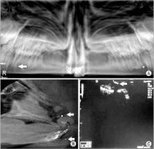
          In this cross-sectional study, five different materials, including wood, metal, glass, tooth and stone, were prepared in three sizes (0.5, 1, and 2 mm) and placed in three locations (soft tissue, air-filled space and bone surface) within a sheep's head (one day after death) and scanned by panoramic radiography, cone-beam computed tomography (CBCT), and ultrasonography (US) devices. The images were reviewed, and accuracy of the detection modalities was recorded. The data were analyzed statistically using the Kruskal-Wallis, Mann-Whitney U-test, Friedman, Wilcoxon signed-rank and kappa tests ( P<0.05).

          Results

          CBCT was more accurate in detection of FBs than panoramic radiography and US ( P<0.001). Metal was the most visible FB in all of modalities. US was the most accurate technique for detecting wooden materials, and CBCT was the best modality for detecting all other materials, regardless of size or location ( P<0.05). The detection accuracy of US was greater in soft tissue, while both CBCT and panoramic radiography had minimal accuracy in detection of FBs in soft tissue.

          Conclusion

          CBCT was the most accurate detection modality for all the sizes, locations and compositions of FBs, except for the wooden materials. Therefore, we recommend CBCT as the gold standard of imaging for detecting FBs in the maxillofacial region.

          Related collections

          Most cited references20

          • Record: found
          • Abstract: found
          • Article: not found

          Diagnosis and treatment of retained foreign bodies in the hand.

          A retrospective review was conducted of 200 consecutive patients with foreign bodies in the hand seen between 1976 and 1982. Wood, glass, and metal accounted for 95 percent of the foreign bodies seen. The duration of follow-up ranged from 1 week to 3 years (average 6 weeks). Approximately 70 percent of the foreign bodies could be removed in either the office or the emergency room. The foreign bodies were removed anywhere from the day of injury to 20 years later. In 38 percent of the patients the diagnosis was missed by the initial treating physician, in many cases because a roentgenography of the injured area was not taken. Metal was visible in all of the radiographic studies obtained, glass in 96 percent, and wood in just 15 percent.
            Bookmark
            • Record: found
            • Abstract: found
            • Article: not found

            Comparison of the sensitivity for detecting foreign bodies among conventional plain radiography, computed tomography and ultrasonography.

            The purpose of this study was to compare the sensitivity for detecting foreign bodies among conventional plain radiography, CT and ultrasonography in in vitro models. Seven different materials were selected as foreign bodies with dimensions of approximately 1 x 1 x 0.1 cm. These materials were metal, glass, wood, stone, acrylic, graphite and Bakelite. These foreign bodies were placed into a sheep's head between the corpus mandible and muscle, in the tongue and in the maxillary sinus. Conventional plain radiography, CT and ultrasonography imaging methods were compared to investigate their sensitivity for detecting these foreign bodies. Metal, glass and stone can be detected with all the visualization techniques used in the study in all of the zones. In contrast to this, foreign bodies with low radiopacity, which could be detected in air with CT, became less visible or almost invisible in muscle tissue and between bone and muscle tissue. The performance of ultrasonography for visualizing foreign bodies with low radiopacity is relatively better than CT. Ultrasonography detects and localizes superficial foreign bodies with low radiopacity in the tissues of the body more effectively than CT and conventional plain radiography. However, CT is a more effective technique for visualization of foreign bodies in air than ultrasound and conventional plain radiography.
              Bookmark
              • Record: found
              • Abstract: found
              • Article: not found

              Wooden foreign bodies in facial injury: a radiological pitfall.

              Foreign bodies can present a diagnostic challenge to the maxillofacial surgeon. Three patients, who suffered from a penetrating injury with a wooden foreign body, were examined and treated. Their preoperative CT and MRI scans were evaluated. In an acute case, the penetrating wooden body mimicked air bubbles. In the other two patients, the wood was retained for several months and appeared with a much higher density on CT. In MRI the wooden foreign bodies gave a low signal intensity. In all injuries removal of the foreign body was delayed, because it was initially radiologically missed or misdiagnosed. In the appropriate trauma setting a penetrating wooden body must always be considered. Its attenuation value increases with time as water is absorbed from the surrounding tissues. Although the radiological appearance may show a great variety, CT imaging is the basic diagnostic technique. MRI is the method of second choice.
                Bookmark

                Author and article information

                Journal
                J Korean Assoc Oral Maxillofac Surg
                J Korean Assoc Oral Maxillofac Surg
                JKAOMS
                Journal of the Korean Association of Oral and Maxillofacial Surgeons
                The Korean Association of Oral and Maxillofacial Surgeons
                2234-7550
                2234-5930
                February 2018
                26 February 2018
                : 44
                : 1
                : 18-24
                Affiliations
                [1 ]Dental Implants Research Center, School of Dentistry, Isfahan University of Medical Science, Isfahan, Iran.
                [2 ]Department of Radiology, School of Dentistry, Isfahan University of Medical Science, Isfahan, Iran.
                [3 ]Department of Oral and Maxillo-Facial Surgery, School of Dentistry, Islamic Azad University, Isfahan (Khorasgan) Branch, Isfahan, Iran.
                Author notes
                Corresponding author: Maedeh Aminian. Department of Radiology, School of Dentistry, Isfahan University of Medical Science, P.O. Box 319, Hezar-Jerib Ave., Isfahan 81746-73461, Iran. TEL: +98-3136680048, FAX: +98-3136699357, maedeh_aminian@ 123456yahoo.com
                Author information
                https://orcid.org/0000-0001-8619-9337
                https://orcid.org/0000-0003-1087-5209
                https://orcid.org/0000-0001-5263-2542
                Article
                10.5125/jkaoms.2018.44.1.18
                5845963
                29535965
                b5618a9a-44a3-48d6-acc6-d7c3743529e9
                Copyright © 2018 The Korean Association of Oral and Maxillofacial Surgeons.

                This is an open-access article distributed under the terms of the Creative Commons Attribution Non-Commercial License ( http://creativecommons.org/licenses/by-nc/4.0/) which permits unrestricted non-commercial use, distribution, and reproduction in any medium, provided the original work is properly cited.

                History
                : 15 July 2017
                : 16 August 2017
                : 16 August 2017
                Categories
                Original Article

                foreign bodies,panoramic radiography,cone-beam computed tomography,ultrasonography,maxillofacial region

                Comments

                Comment on this article