7
views
0
recommends
+1 Recommend
0 collections
    0
    shares
      • Record: found
      • Abstract: found
      • Article: found
      Is Open Access

      Single cell RNA-seq analysis identifies ferroptotic chondrocyte cluster and reveals TRPV1 as an anti-ferroptotic target in osteoarthritis

      research-article

      Read this article at

      Bookmark
          There is no author summary for this article yet. Authors can add summaries to their articles on ScienceOpen to make them more accessible to a non-specialist audience.

          Summary

          Background

          Osteoarthritis (OA) is the most common degenerative joint disease primarily characterized by cartilage destruction. The aim of this study was to investigate the role, molecular characteristics and potential therapeutic target of chondrocyte ferroptosis in the pathogenesis of OA.

          Methods

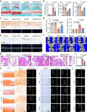
          The expression of ferroptotic hallmarks (iron and lipid peroxidation accumulation, glutathione deletion) were analyzed in paired intact and damaged cartilages from OA patients. Single cell RNA sequencing (scRNA-seq) analysis was performed on 17,638 chondrocytes to verify the presence, investigate the molecular signatures and unveil the potential therapeutic target of ferroptotic chondrocyte cluster in human OA cartilages. Destabilization of medial meniscus (DMM)-induced OA model and tert-butyl hydroperoxide (TBHP)-treated primary mouse chondrocytes and human cartilage explants were used to evaluate the protective effect of pharmacologically activated transient receptor potential vanilloid 1 (TRPV1). The downstream molecular mechanisms of TRPV1 was further investigated in glutathione peroxidase 4 (Gpx4) heterozygous genetic deletion mice ( Gpx4 +/− ).

          Findings

          The concentrations of iron and lipid peroxidation and the expression of ferroptotic drivers in the damaged areas of human OA cartilages were significantly higher than those in the intact cartilage. scRNA-seq analysis revealed a chondrocyte cluster characterized by preferentially expressed ferroptotic hallmarks and genes, namely ferroptotic chondrocyte cluster. Comprehensive gene set variation analysis revealed TRPV1 as an anti-ferroptotic target in human OA cartilage. Pharmacological activation of TRPV1 significantly abrogated cartilage degeneration by protecting chondrocytes from ferroptosis. Mechanistically, TRPV1 promoted the expression of GPX4, and its anti-ferroptotic role was largely mitigated in the OA model of Gpx4 +/− mice.

          Interpretation

          TRPV1 activation protects chondrocytes from ferroptosis and ameliorates OA progression by upregulating GPX4.

          Funding

          National Key R&D Program of China (2018YFC1105904), Key Program of NSFC (81730067), National Science Foundation of China (81772335, 81941009, 81802196), Natural Science Foundation of Jiangsu Province, China (BK20180127), Jiangsu Provincial Key Medical Talent Foundation, Six Talent Peaks Project of Jiangsu Province (WSW-079).

          Related collections

          Most cited references58

          • Record: found
          • Abstract: found
          • Article: not found

          Ferroptosis: an iron-dependent form of nonapoptotic cell death.

          Nonapoptotic forms of cell death may facilitate the selective elimination of some tumor cells or be activated in specific pathological states. The oncogenic RAS-selective lethal small molecule erastin triggers a unique iron-dependent form of nonapoptotic cell death that we term ferroptosis. Ferroptosis is dependent upon intracellular iron, but not other metals, and is morphologically, biochemically, and genetically distinct from apoptosis, necrosis, and autophagy. We identify the small molecule ferrostatin-1 as a potent inhibitor of ferroptosis in cancer cells and glutamate-induced cell death in organotypic rat brain slices, suggesting similarities between these two processes. Indeed, erastin, like glutamate, inhibits cystine uptake by the cystine/glutamate antiporter (system x(c)(-)), creating a void in the antioxidant defenses of the cell and ultimately leading to iron-dependent, oxidative death. Thus, activation of ferroptosis results in the nonapoptotic destruction of certain cancer cells, whereas inhibition of this process may protect organisms from neurodegeneration. Copyright © 2012 Elsevier Inc. All rights reserved.
            Bookmark
            • Record: found
            • Abstract: found
            • Article: not found

            Ferroptosis: mechanisms, biology and role in disease

            The research field of ferroptosis has seen exponential growth over the past few years, since the term was coined in 2012. This unique modality of cell death, driven by iron-dependent phospholipid peroxidation, is regulated by multiple cellular metabolic pathways, including redox homeostasis, iron handling, mitochondrial activity and metabolism of amino acids, lipids and sugars, in addition to various signalling pathways relevant to disease. Numerous organ injuries and degenerative pathologies are driven by ferroptosis. Intriguingly, therapy-resistant cancer cells, particularly those in the mesenchymal state and prone to metastasis, are exquisitely vulnerable to ferroptosis. As such, pharmacological modulation of ferroptosis, via both its induction and its inhibition, holds great potential for the treatment of drug-resistant cancers, ischaemic organ injuries and other degenerative diseases linked to extensive lipid peroxidation. In this Review, we provide a critical analysis of the current molecular mechanisms and regulatory networks of ferroptosis, the potential physiological functions of ferroptosis in tumour suppression and immune surveillance, and its pathological roles, together with a potential for therapeutic targeting. Importantly, as in all rapidly evolving research areas, challenges exist due to misconceptions and inappropriate experimental methods. This Review also aims to address these issues and to provide practical guidelines for enhancing reproducibility and reliability in studies of ferroptosis. Finally, we discuss important concepts and pressing questions that should be the focus of future ferroptosis research.
              Bookmark
              • Record: found
              • Abstract: not found
              • Article: not found

              FSP1 is a glutathione-independent ferroptosis suppressor

                Bookmark

                Author and article information

                Contributors
                Journal
                eBioMedicine
                EBioMedicine
                eBioMedicine
                Elsevier
                2352-3964
                19 September 2022
                October 2022
                19 September 2022
                : 84
                : 104258
                Affiliations
                [a ]State Key Laboratory of Pharmaceutical Biotechnology, Division of Sports Medicine and Adult Reconstructive Surgery, Department of Orthopedic Surgery, Nanjing Drum Tower Hospital, The Affiliated Hospital of Nanjing University Medical School, Nanjing 210008, Jiangsu, PR China
                [b ]Dr. Li Dak Sum-Yip Yio Chin Center for Stem Cells and Regenerative Medicine and Department of Orthopedic Surgery of The Second Affiliated Hospital, Zhejiang University School of Medicine, Hangzhou 310000, PR China
                [c ]Key Laboratory of Tissue Engineering and Regenerative Medicine of Zhejiang Province, Zhejiang University School of Medicine, Hangzhou 310000, PR China
                [d ]Nanjing Drum Tower Hospital Clinical College of Traditional Chinese and Western Medicine, Nanjing University of Chinese Medicine, Nanjing 210008, Jiangsu, PR China
                [e ]Department of orthopedics, The Affiliated Jiangyin Hospital of Southeast University Medical College, Wuxi, Jiangsu 214400, PR China
                [f ]Laboratory for Bone and Joint Diseases, RIKEN Center for Integrative Medical Science (IMS, RIKEN), Tokyo 108-8639, Japan
                [g ]Department of Sports Medicine, Zhejiang University School of Medicine, Hangzhou 310000, PR China
                [h ]China Orthopedic Regenerative Medicine Group (CORMed), Hangzhou 310000, PR China
                Author notes
                [* ]Corresponding author at: Dr. Li Dak Sum-Yip Yio Chin Center for Stem Cells and Regenerative Medicine and Department of Orthopedic Surgery of The Second Affiliated Hospital, Zhejiang University School of Medicine, Hangzhou 310000, PR China. chenxiao-610@ 123456zju.edu.cn
                [** ]Corresponding author at: State Key Laboratory of Pharmaceutical Biotechnology, Division of Sports Medicine and Adult Reconstructive Surgery, Department of Orthopedic Surgery, Nanjing Drum Tower Hospital, The Affiliated Hospital of Nanjing University Medical School, Nanjing 210008, Jiangsu, PR China shidongquan@ 123456nju.edu.cn
                [1]

                Zhongyang Lv, Jie Han and Jiawei Li contributed equally to this work.

                Article
                S2352-3964(22)00440-6 104258
                10.1016/j.ebiom.2022.104258
                9494174
                36137413
                b50c3ef3-3d03-40a8-9f10-86002a3fec0d
                © 2022 The Author(s)

                This is an open access article under the CC BY-NC-ND license (http://creativecommons.org/licenses/by-nc-nd/4.0/).

                History
                : 15 April 2022
                : 25 August 2022
                : 25 August 2022
                Categories
                Articles

                osteoarthritis,ferroptosis,single cell rna sequencing,trpv1,gpx4

                Comments

                Comment on this article