0
views
0
recommends
+1 Recommend
0 collections
    0
    shares
      • Record: found
      • Abstract: found
      • Article: found
      Is Open Access

      Dislocated Intraocular Lens Extraction and Iris-Claw Lens Implantation in Vitrectomized and Non-vitrectomized Eyes

      research-article

      Read this article at

      Bookmark
          There is no author summary for this article yet. Authors can add summaries to their articles on ScienceOpen to make them more accessible to a non-specialist audience.

          Abstract

          Objectives:

          To compare the outcomes and complications of dislocated intraocular lens (IOL) extraction and secondary iris-claw IOL (ICIOL) implantation in vitrectomized and non-vitrectomized eyes.

          Materials and Methods:

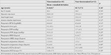
          This retrospective study included 19 vitrectomized eyes and 11 non-vitrectomized eyes that underwent dislocated IOL extraction and secondary anterior chamber ICIOL implantation between June 2014 and September 2017 and had at least one year of follow-up.

          Results:

          There were no significant differences between the groups in terms of demographic data, operative time, baseline anatomic and functional measurements, or postoperative changes in these measurements (all p>0.05). Postoperative best corrected visual acuity was significantly higher than preoperative values in both groups (both p<0.05). Complication rates did not differ between the groups (all p>0.05). In both groups, endothelial cell density was significantly lower at postoperative 1 year compared to preoperative measurements. There was no significant difference between groups regarding endothelial cell loss (p=0.49). One vitrectomized eye had corneal decompensation. Other complications included hyphema, transient increase of intraocular pressure, secondary glaucoma, pupillary irregularity, and dislocation of ICIOL. Mean operative time was 26.4±5.9 minutes.

          Conclusion:

          Dislocated IOL extraction and secondary anterior chamber ICIOL implantation is a safe treatment option in both vitrectomized and non-vitrectomized eyes.

          Related collections

          Most cited references25

          • Record: found
          • Abstract: found
          • Article: not found

          Iris-claw intraocular lenses to correct aphakia in the absence of capsule support.

          To evaluate the indications, postoperative visual efficacy, and complication rate after intraocular implantation of an iris-claw aphakic intraocular lens (IOL). Oxford Eye Hospital, Oxford, United Kingdom. Case series. This chart review comprised eyes with no capsule support that had anterior iris-fixation IOL implantation for aphakia between 2001 and 2009. The study comprised 116 eyes (104 patients). Iris-claw IOLs were inserted during primary lens surgery in 18 eyes (15.5%), during an IOL exchange procedure for dislocated posterior chamber IOLs in 19 eyes (16.4%), and as a secondary procedure in 79 eyes (68.1%). The mean follow-up was 22.4 months (range 3 to 79 months). The final corrected distance visual acuity (CDVA) was 6/12 or better in 68.9% of all eyes and in 47 of 53 eyes (88.7%) with no preoperative comorbidity. Complications included wound leak requiring resuturing in 2.6% of eyes, postoperative intraocular pressure rise in 9.5% of eyes (glaucoma escalation 0.8%), and cystoid macular edema in 7.7% of eyes (0.8% chronic). Iris-claw IOL subluxation occurred in 6.0% of eyes from 5 days to 60 months postoperatively; all the IOLs were repositioned. Corneal decompensation occurred in 1.7% of eyes; 0.8% had retinal detachments. Iris-claw IOL implantation for aphakia gave a good visual outcome and can be used for a wide range of indications. Postoperative complication rates were comparable to, if not better than, those with conventional anterior chamber IOLs. Correct implantation technique is critical in avoiding postoperative IOL subluxation. Copyright © 2011 ASCRS and ESCRS. Published by Elsevier Inc. All rights reserved.
            Bookmark
            • Record: found
            • Abstract: found
            • Article: not found

            Posterior iris fixation of the iris-claw intraocular lens implantation through a scleral tunnel incision.

            To evaluate the technique, efficacy, and safety of posterior iris fixation of iris-claw intraocular lens (IOLs) implantation through a scleral tunnel incision for aphakia correction. Noncomparative, interventional case series. A secondary posterior iris fixation of the Artisan iris-claw IOL (Ophthec BV, Groningen, The Netherlands) was implanted for aphakia correction in the authors' clinical practice. Uncorrected visual acuity, best spectacle-corrected visual acuity (BSCVA), astigmatism, manifest refraction, lens position, pigment dispersion, and intraocular pressure (IOP) were evaluated in 32 consecutive eyes of 32 patients. BSCVA was 20/40 or better in 28 eyes (87.50%) during the mean follow-up time (nine months). Mean postoperative spherical equivalent was -0.70 diopters (D; standard deviation [SD], 0.47 D) at six months after surgery. Mean prediction error was -0.13 D (SD, 0.28 D), and mean absolute prediction error was 0.26 D (SD, 0.15 D). Preoperative mean astigmatism was -1.08 D (SD, 0.55 D; range, 0.0 to -2.0 D). At six months after surgery, mean astigmatism was -2.1 D (SD, 0.81 D; range, -0.75 to -3.75 D). There was no significant postoperative IOP increase. Lens position, evaluated by Oculus Pentacam (Pentacam 70700: Oculus, Wetzlar, Germany) and ultrasound biomicroscopy [UBM] (Ophthalmic Technologies Inc, Toronto, Ontario, Canada), was parallel to the iris plane. Posterior iris fixation of the iris-claw IOL implantation through a scleral tunnel incision is a safe procedure and an effective option for aphakic eyes without capsule support.
              Bookmark
              • Record: found
              • Abstract: found
              • Article: not found

              Lens implant selection with absence of capsular support.

              If contact lens or spectacle correction is not viable, little debate exists that the secondary placement of an intra-ocular lens (IOL) is the method of choice in the absence of capsular support. The choice of IOL mainly depends on the preoperative status of the eye (eg, aphakia in children) and the selected location for the implant. Theoretically, there are several IOL implantation approaches in cases without capsular support: an angle-supported anterior chamber (AC) IOL, an iris-fixated ACIOL, an iris-sutured or iris-fixated posterior chamber (PC) IOL and a transsclerally sutured PCIOL. No consensus exists, however, on the indications as well as on the relative safety and efficacy of these different options. Implantation of modern ACIOLs, like the refined open-loop or iris-fixated claw (toric) ACIOLs, have regained popularity and provide a valuable alternative to sutured PCIOLs. However, in the absence of capsular support, the transsclerally sutured PCIOLs offer numerous advantages for certain eyes. Because of its anatomic location, the sutured PCIOL is more appropriate for eyes with compromised cornea, peripheral anterior synechiae, shallow anterior chamber, or glaucoma. Moreover, sutured PCIOLs are appropriate if the patient with aphakia is young or has a life expectancy of 10 years or more. Recent technological advances, including PCIOL with iris diaphragm for aniridia, toric ACIOLs, and small-incision surgery with foldable, transsclerally sutured IOLs, seem to further improve clinical outcomes.
                Bookmark

                Author and article information

                Journal
                Turk J Ophthalmol
                Turk J Ophthalmol
                TJO
                Turkish Journal of Ophthalmology
                Galenos Publishing
                2149-8695
                2149-8709
                October 2019
                24 October 2019
                : 49
                : 5
                : 277-282
                Affiliations
                [1 ]İstanbul Retina Institute, İstanbul, Turkey
                Author notes
                * Address for Correspondence: İstanbul Retina Institute, İstanbul, Turkey Phone: +90 212 231 31 21 E-mail: mkaracorlu@ 123456gmail.com
                Author information
                https://orcid.org/0000-0002-2336-0696
                https://orcid.org/0000-0002-8081-0032
                https://orcid.org/0000-0002-5464-8713
                https://orcid.org/0000-0002-3430-4868
                https://orcid.org/0000-0001-5561-3339
                Article
                30596
                10.4274/tjo.galenos.2019.79735
                6823582
                31650810
                b32fa230-8ae3-4f9a-bee7-778453fdeb0f
                © Copyright 2019 by Turkish Ophthalmological Association | Turkish Journal of Ophthalmology, published by Galenos Publishing House.

                This is an open-access article distributed under the terms of the Creative Commons Attribution License, which permits unrestricted use, distribution, and reproduction in any medium, provided the original work is properly cited.

                History
                : 17 December 2018
                : 6 May 2019
                Categories
                Original Article

                aphakia,intraocular lens dislocation,iris-claw lens,secondary lens implantation,vitrectomy

                Comments

                Comment on this article