0
views
0
recommends
+1 Recommend
0 collections
    0
    shares
      • Record: found
      • Abstract: found
      • Article: found
      Is Open Access

      Morphological Analysis of the Sylvian Fissure Stem to Guide a Safe Trans-sylvian Fissure Approach

      research-article

      Read this article at

      Bookmark
          There is no author summary for this article yet. Authors can add summaries to their articles on ScienceOpen to make them more accessible to a non-specialist audience.

          Abstract

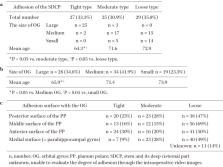
          The sylvian fissure stem and its deep cisternal part (SDCP) consist mainly of the orbital gyrus (OG) and anterior medial portion of the temporal lobe. SDCP's adhesion has been found to make a trans-sylvian approach difficult due to the various patterns of adhesion. Thus, in this study, we aim to clarify the morphological features of the SDCP, and to guide a safe trans-sylvian approach. We retrospectively classified the morphology of the SDCP in 81 patients into 3 types (tight, moderate, loose type) according to the degree of adhesion of the arachnoid membrane and analyzed the morphological features of the OG and the temporal lobe using intraoperative video images. In addition, we have retrospectively measured each width of the SDCP's subarachnoid space at the three points (Point A, lateral superior portion; Point B, downward portion; Point C, medial inferior portion of SDCP) and analyzed their relationship to the degree of adhesion using the preoperative coronal three-dimensional computed tomography angiography (3D-CTA) images of 44 patients. As per the results, SDCP's adhesions were determined to be significantly tighter in cases with large OG and young cases. The temporal lobe had four surfaces (posterior, middle, anterior, and medial) that adhered to the OG in various patterns. The tighter the adhesion between the OG and each of the three distal surfaces of the temporal lobe, the narrower the width of the subarachnoid space at each point (A, B, C). Understanding of the morphological features of the SDCP, and estimating its adhesion preoperatively are useful in developing a surgical strategy and obtaining correct intraoperative orientation in the trans-sylvian approach.

          Related collections

          Most cited references37

          • Record: found
          • Abstract: found
          • Article: not found

          Microsurgical anatomy of the middle cerebral artery.

          The microsurgical anatomy of the middle cerebral artery (MCA) was defined in 50 cerebral hemispheres. The MCA was divided into four segments: the M1 (sphenoidal) segment coursed posterior and parallel to the sphenoid ridge; the M2 (insular) segment lay on the insula; the M3 (opercular) segment coursed over the frontoparietal and temporal opercula; and the M4 (cortical) segment spread over the cortical surface. The Sylvian fissure was divided into a sphenoidal and an operculoinsular compartment. The M1 segment coursed in the sphenoidal compartment, and the M2 and M3 segments coursed in the operculoinsular compartment. The main trunk of the MCA divided in one of three ways; bifurcation (78% of hemispheres), trifurcation (12%), or division into multiple trunks (10%). The MCA's that bifurcated were divided into three groups: equal bifurcation (18%), inferior trunk dominant (32%), or superior trunk dominant (28%). The MCA territory was divided into 12 areas: orbitofrontal, prefrontal, precentral, central, anterior parietal, posterior parietal, angular, temporo-occipital, posterior temporal, middle temporal, anterior temporal, and temporopolar. The smallest cortical arteries arose at the anterior end and the largest one at the posterior end of the Sylvian fissure. The largest cortical arteries supplied the temporo-occipital and angular areas. The relationship of each of the cortical arteries to a number of external landmarks was reviewed in detail.
            Bookmark
            • Record: found
            • Abstract: found
            • Article: not found

            Topographic anatomy of the insular region.

            The insula is one of the paralimbic structures and constitutes the invaginated portion of the cerebral cortex, forming the base of the sylvian fissure. The authors provide a detailed anatomical study of the insular region to assist in the process of conceptualizing a reliable surgical approach to allow for a successful course of surgery. The topographic anatomy of the insular region was studied in 25 formalin-fixed brain specimens (50 hemispheres). The periinsular sulci (anterior, superior, and inferior) define the limits of the frontoorbital, frontoparietal, and temporal opercula, respectively. The opercula cover and enclose the insula. The limen insula is located in the depths of the sylvian fissure and constitutes the anterobasal portion of the insula. A central insular sulcus divides the insula into two portions, the anterior insula (larger) and the posterior insula (smaller). The anterior insula is composed of three principal short insular gyri (anterior, middle, and posterior) as well as the accessory and transverse insular gyri. All five gyri converge at the insular apex, which represents the most superficial aspect of the insula. The posterior insula is composed of the anterior and posterior long insular gyri and the postcentral insular sulcus, which separates them. The anterior insula was found to be connected exclusively to the frontal lobe, whereas the posterior insula was connected to both the parietal and temporal lobes. Opercular gyri and sulci were observed to interdigitate within the opercula and to interdigitate the gyri and sulci of the insula. Using the fiber dissection technique, various unique anatomical features and relationships of the insula were determined. The topographic anatomy of the insular region is described in this article, and a practical terminology for gyral and sulcal patterns of surgical significance is presented. This study clarifies and supplements the information presently available to help develop a more coherent surgical concept.
              Bookmark
              • Record: found
              • Abstract: found
              • Article: not found

              Microsurgical pterional approach to aneurysms of the basilar bifurcation.

              The authors describe a microsurgical frontolateral pterional approach to aneurysms of the basilar bifurcation. Results of surgery in 38 patients are presented.
                Bookmark

                Author and article information

                Journal
                Neurol Med Chir (Tokyo)
                Neurol Med Chir (Tokyo)
                Neurologia medico-chirurgica
                The Japan Neurosurgical Society
                0470-8105
                1349-8029
                22 September 2022
                November 2022
                : 62
                : 11
                : 502-512
                Affiliations
                [1 ]Department of Neurosurgery, Yamada Memorial Hospital, Mihara, Hiroshima, Japan
                [2 ]Department of Neurosurgery, Chugoku Rosai Hospital, Kure, Hiroshima, Japan
                [3 ]Department of Neurosurgery, Graduate School of Biomedical and Health Sciences, Hiroshima University, Hiroshima, Hiroshima, Japan
                Author notes

                Corresponding author: Yasutaka Imada, MD., PhD.

                Department of Neurosurgery, Yamada Memorial Hospital, 6-2-1 Miyaura, Mihara-shi, Hiroshima 723-0051, Japan.

                e-mail: yasutaka5682@ 123456yahoo.co.jp

                Article
                10.2176/jns-nmc.2022-0064
                9726177
                36130902
                b21f0710-88d8-4d48-8a2c-77fb4a4555be
                © 2022 The Japan Neurosurgical Society

                This work is licensed under a Creative Commons Attribution-NonCommercial-NoDerivatives International License.

                History
                : 1 March 2022
                : 11 July 2022
                Categories
                Original Article

                orbital gyrus,planum polare of the temporal lobe,sylvian fissure stem,trans-sylvian approach

                Comments

                Comment on this article