16
views
0
recommends
+1 Recommend
0 collections
    0
    shares
      • Record: found
      • Abstract: found
      • Article: found
      Is Open Access

      Inverted ILM flap, free ILM flap and conventional ILM peeling for large macular holes

      research-article

      Read this article at

      Bookmark
          There is no author summary for this article yet. Authors can add summaries to their articles on ScienceOpen to make them more accessible to a non-specialist audience.

          Abstract

          Background

          To assess closure rate after a single surgery of large macular holes and their visual recovery in the short term with three different surgical techniques.

          Methods

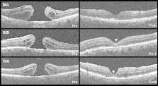
          Prospective multicenter randomized controlled trial. We included treatment-naïve patients with diagnosis of large macular hole (minimum diameter of > 400 µm). All patients underwent a comprehensive ophthalmological examination. Before surgery, the patients were randomized into three groups: group A: conventional internal limiting membrane peeling, group B: inverted-flap technique and group C: free-flap technique. All study measurements were repeated within the period of 1 and 3 months after surgery. Continuous variables were assessed with a Kruskal–Wallis test, change in visual acuity was assessed with analysis of variance for repeated measurements with a Bonferroni correction for statistical significance.

          Results

          Thirty-eight patients were enrolled (group A: 12, group B: 12, group C: 14). The closure rate was in group A and B: 91.6%; 95% CI 61.52–99.79%. In group C: 85.71%; 95% CI 57.19–98.22%. There were no differences in the macular hole closure rate between groups ( p = 0.85). All groups improved ≈ 0.2 logMAR, but only group B reached statistical significance ( p < 0.007).

          Conclusions

          Despite all techniques displayed a trend toward visual improvement, the inverted-flap technique seems to induce a faster and more significant recovery in the short term.

          Related collections

          Most cited references40

          • Record: found
          • Abstract: found
          • Article: not found

          Inverted internal limiting membrane flap technique for large macular holes.

          Large macular holes usually have an increased risk of surgical failure. Up to 44% of large macular holes remain open after 1 surgery. Another 19% to 39% of macular holes are flat-open after surgery. Flat-open macular holes are associated with limited visual acuity. This article presents a modification of the standard macular hole surgery to improve functional and anatomic outcomes in patients with large macular holes. A prospective, randomized clinical trial. Patients with macular holes larger than 400 μm were included. In group 1, 51 eyes of 40 patients underwent standard 3-port pars plana vitrectomy with air. In group 2, 50 eyes of 46 patients underwent a modification of the standard technique, called the inverted internal limiting membrane (ILM) flap technique. In the inverted ILM flap technique, instead of completely removing the ILM after trypan blue staining, a remnant attached to the margins of the macular hole was left in place. This ILM remnant was then inverted upside-down to cover the macular hole. Fluid-air exchange was then performed. Spectral optical coherence tomography and clinical examination were performed before surgery and postoperatively at 1 week and 1, 3, 6, and 12 months. Visual acuity and postoperative macular hole closure. Preoperative mean visual acuity was 0.12 in group 1 and 0.078 in group 2. Macular hole closure was observed in 88% of patients in group 1 and in 98% of patients in group 2. A flat-hole roof with bare retinal pigment epithelium (flat-open) was observed in 19% of patients in group 1 and 2% of patients in group 2. Mean (or median) postoperative visual acuity 12 months after surgery was 0.17 (range, 0.1-0.6) in group 1 and 0.28 (range, 0.02-0.8) in group 2 (P = 0.001). The inverted ILM flap technique prevents the postoperative flat-open appearance of a macular hole and improves both the functional and anatomic outcomes of vitrectomy for macular holes with a diameter greater than 400 μm. Spectral optical coherence tomography after vitrectomy with the inverted ILM flap technique suggests improved foveal anatomy compared with the standard surgery. Copyright © 2010 American Academy of Ophthalmology. Published by Elsevier Inc. All rights reserved.
            Bookmark
            • Record: found
            • Abstract: found
            • Article: not found

            Vitreous surgery for idiopathic macular holes. Results of a pilot study.

            Idiopathic macular holes are generally considered an untreatable condition. We used modern vitrectomy techniques to evaluate two questions: (1) Is it possible to reattach the retina around the macular hole? (2) If it is reattached, will the patient's central vision improve? In 30 (58%) of 52 patients, we were able to reattach successfully the detached macula with our surgical procedure. In 22 (73%) of the 30 patients in whom the macula was successfully reattached, there was an improvement in visual acuity of two lines or better. In the 22 patients in whom reattachment of the macular hole was not obtained, there was no significant improvement in visual acuity. Thus, the overall success rate for improved vision postoperatively was 42% (22/52). Complications related to surgery were observed in eight patients (15%) early in our experience with this procedure and included increase in the size of the macular hole, mottling of the retinal pigmented epithelium, and a vascular occlusion. Our clinical observations indicate that the treatment of macular holes by vitrectomy may offer some promise for this otherwise untreatable condition. In patients in whom reattachment was successful, the technique used appeared to allow for clinically significant improvements in visual acuity. However, additional work on increasing surgical success and minimizing surgical complications, as well as a further understanding of the mechanism of retinal reattachment, is required before widespread use of this procedure for treating macular holes.
              Bookmark
              • Record: found
              • Abstract: found
              • Article: not found

              Autologous transplantation of the internal limiting membrane for refractory macular holes.

              To determine the effectiveness of autologous transplantation of the internal limiting membrane (ILM) for refractory macular holes.
                Bookmark

                Author and article information

                Contributors
                +52.55.1084.1400 , rvelezmx@yahoo.com , rvelezmx@yahoo.mx
                ramirezestudillo@gmail.com
                carlgustaf.sjoholm@gmail.com
                franciscobejar_cornejo@hotmail.com
                jorgesr84@hotmail.com
                guenarmx@yahoo.com.mx
                vmoralesc@mac.com
                tolodamota@yahoo.com.mx
                Journal
                Int J Retina Vitreous
                Int J Retina Vitreous
                International Journal of Retina and Vitreous
                BioMed Central (London )
                2056-9920
                19 February 2018
                19 February 2018
                2018
                : 4
                : 8
                Affiliations
                [1 ]GRID grid.464508.b, Retina Department, , Asociación para Evitar la Ceguera en Mexico, Hospital “Dr. Luis Sanchez Bulnes” IAP, ; Vicente García Torres #46, Col: San Lucas Coyoacán, 04030 Mexico City, DF Mexico
                [2 ]Retina and Vitreous Department, Fundación Hospital Nuestra Señora de la Luz, Mexico City, Mexico
                [3 ]Ophthalmology Unit, Clinica David, Morelia City, Mexico
                Author information
                http://orcid.org/0000-0002-6457-4578
                Article
                111
                10.1186/s40942-018-0111-5
                5817800
                29479478
                b1d2626e-21d2-49ec-814e-9f47d42c1ba6
                © The Author(s) 2018

                Open AccessThis article is distributed under the terms of the Creative Commons Attribution 4.0 International License ( http://creativecommons.org/licenses/by/4.0/), which permits unrestricted use, distribution, and reproduction in any medium, provided you give appropriate credit to the original author(s) and the source, provide a link to the Creative Commons license, and indicate if changes were made. The Creative Commons Public Domain Dedication waiver ( http://creativecommons.org/publicdomain/zero/1.0/) applies to the data made available in this article, unless otherwise stated.

                History
                : 25 September 2017
                : 20 January 2018
                Categories
                Original Article
                Custom metadata
                © The Author(s) 2018

                large macular hole,internal limiting membrane,inverted-flap,free-flap,visual recovery,surgery,treatment

                Comments

                Comment on this article