4
views
0
recommends
+1 Recommend
0 collections
    0
    shares
      • Record: found
      • Abstract: found
      • Article: found
      Is Open Access

      Urethral meatus stricture BOO stimulates bladder smooth muscle cell proliferation and pyroptosis via IL-1β and the SGK1-NFAT2 signaling pathway

      research-article

      Read this article at

      Bookmark
          There is no author summary for this article yet. Authors can add summaries to their articles on ScienceOpen to make them more accessible to a non-specialist audience.

          Abstract

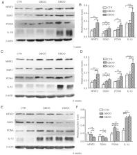
          Bladder outlet obstruction (BOO), which is primarily caused by benign prostatic hyperplasia, is a common chronic disease. However, previous studies have most commonly investigated BOO using the acute obstruction model. In the present study, a chronic obstruction model was established to investigate the different pathological alterations in the bladder between acute and chronic obstruction. Compared with chronic obstruction, acute obstruction led to increased expression of proliferating cell nuclear antigen and interleukin-1β, which are markers of proliferation and inflammation, respectively. Furthermore, increased fibrosis in the bladder at week 2 was observed. Low pressure promoted mice bladder smooth muscle cell (MBSMC) proliferation, and pressure overload inhibited cell proliferation and increased the proportion of dead MBSMCs. Further investigation using serum/glucocorticoid regulated kinase 1 (SGK1) small interfering RNAs indicated that low pressure may promote MBSMC proliferation by upregulating SGK1 and nuclear factor of activated T-cell expression levels. Therefore, the present study suggested that acute obstruction led to faster decompensation of bladder function and chronic bladder obstruction displayed an enhanced ability to progress to BOO.

          Related collections

          Most cited references46

          • Record: found
          • Abstract: found
          • Article: not found

          Prognostic value of urodynamic testing in myelodysplastic patients.

          We herein describe the clinical progress of 42 myelodysplastic patients studied urodynamically and followed for a mean of 7.1 years. Urodynamic evaluation included urethral pressure profilometry, simultaneous determination of urethral pressure, intravesical pressure and external anal or external urethral sphincter electromyography with fluoroscopic voiding cystourethrography. Assessment of urethral function showed 36 patients (86 per cent) with an open vesical outlet and nonfunctional proximal urethral. Cystometrography revealed that 7 of 42 patients (17 per cent) had reflex detrusor activity: 4 with coordinated micturition and 3 with detrusor-sphincter dyssynergia. Thirty-five patients (83 per cent) had areflexic detrusor dysfunction: 5 with atomic detrusor response and 30 with a progressive increase in pressure with increasing volume. The intravesical pressure at the time of urethral leakage was 40 cm. water or less in 20 patients and at pressures greater than this value in 22 patients. No patient in the low pressure group had vesicoureteral reflux and only 2 showed ureteral dilatation on excretory urography. In contrast, of the patients in the higher pressure group 15 (68 per cent) showed vesicoureteral reflux and 18 (81 per cent) showed ureteral dilatation on excretory urography. Thus, a striking relationship between the urethral closure pressure and intravesical pressure at the time of urethral leakage and the clinical course in this group of myelodysplastic patients is demonstrated. Every patient with a normally closed vesical outlet was continent on intermittent catheterization and an anticholinergic agent, while only 60 per cent of patients with open bladder outlets similarly treated achieved good urinary control and none was dry. An artificial sphincter device would seem to be a reasonable method to achieve urinary control in the latter patients but the detrusor response to filling also must be considered. Detrusor hypertonia should be controlled or controllable before a sphincter augmenting device can be used safely. Treatment options for patients with high urethral closure pressures include intermittent catheterization and anticholinergic medications or a sphincter ablative procedure to decrease the outlet resistance combined with anticholinergic therapy and implantation of an artificial sphincter. However, only longer followup will determine if these therapeutic regimens will prevent upper urinary tract deterioration.
            Bookmark
            • Record: found
            • Abstract: found
            • Article: found
            Is Open Access

            Cytokine Secretion and Pyroptosis of Thyroid Follicular Cells Mediated by Enhanced NLRP3, NLRP1, NLRC4, and AIM2 Inflammasomes Are Associated With Autoimmune Thyroiditis

            Background Inflammasomes, which mediate maturation of interleukin-1β (IL-β) and interleukin-18 (IL-18) and lead to pyroptosis, have been linked to various autoimmune disorders. This study investigated whether they are involved in the pathogenesis of autoimmune thyroiditis (AIT). Methods We collected thyroid tissues from 50 patients with AIT and 50 sex- and age-matched controls. Serum levels of free T3, free T4, thyrotropin, thyroid peroxidase antibody (TPOAb), and thyroglobulin antibody (TgAb) were measured by electrochemiluminescent immunoassays. Expression of several inflammasome components, the NOD-like receptor (NLR) family pyrin domain containing 1 (NLRP1), NLRP3, CARD-domain containing 4 (NLRC4), absent in melanoma 2 (AIM2), the apoptosis-associated speck-like protein that contains a caspase recruitment domain (ASC), caspase-1, IL-1β, and IL-18 was determined by real-time PCR and western blot. Immunohistochemistry was used to localize the expression of NLRP1, NLRP3, NLRC4, and AIM2. The Nthy-ori 3-1 thyroid cell line was stimulated with tumor necrosis factor-α (TNF-α), interferon-γ (IFN-γ), interleukin-17A, interleukin-6, and poly(dA:dT). The levels of IL-18 and IL-1β in the cell supernatant were measured by enzyme-linked immunosorbent assay, and lactate dehydrogenase was quantified by absorptiometry. ASC specks were examined by confocal immunofluorescence microscopic analysis. Cell death was examined by flow cytometry, and the N-terminal domain of gasdermin D was detected by western blot analysis. Results Expression of NLRP1, NLRP3, NLRC4, AIM2, ASC, caspase-1, pro IL-1β, pro IL-18, mRNA, and protein was significantly increased in thyroid tissues from patients with AIT, and enhanced posttranslational maturation of caspase-1, IL-18 and IL-1β was also observed. Expression of NLRP1, NLRP3, NLRC4, and AIM2 was localized mainly in thyroid follicular cells adjacent to areas of lymphatic infiltration. The thyroid mRNA level of NLRP1 and ASC was correlated to the serum TPOAb and TgAb levels in the AIT group. TNF-α and IFN-γ had a priming effect on the expression of multiple inflammasome components in thyroid cells. IFN-γ was found to strengthen poly(dA:dT)-induced cell pyroptosis and bioactive IL-18 release. Conclusion Our work has demonstrated for the first time that multiple inflammasomes are associated with AIT pathogenesis. The identified NLRP3, NLRP1, NLRC4, AIM2 inflammasomes and their downstream cytokines may represent potential therapeutic targets and biomarkers of AIT.
              Bookmark
              • Record: found
              • Abstract: found
              • Article: found
              Is Open Access

              Progressive bladder remodeling due to bladder outlet obstruction: a systematic review of morphological and molecular evidences in humans

              Background Bladder outlet obstruction is a common urological condition. We aimed to summarize available evidences about bladder outlet obstruction-induced molecular and morphological alterations occurring in human bladder. Methods We performed a literature search up to December 2017 including clinical and preclinical basic research studies on humans. The following search terms were combined: angiogenesis, apoptosis, bladder outlet obstruction, collagen, electron microscopy, extracellular matrix, fibrosis, hypoxia, histology, inflammation, innervation, ischemia, pressure, proliferation, remodeling, suburothelium, smooth muscle cells, stretch, urothelium. Results We identified 36 relevant studies. A three-stages model of bladder wall remodeling can be hypothesized involving an initial hypertrophy phase, a subsequent compensation phase and a later decompensation. Histological and molecular alterations occur in the following compartments: urothelium, suburothelium, detrusor smooth muscle cells, detrusor extracellular matrix, nerves. Cyclic stretch, increased hydrostatic and cyclic hydrodynamic pressure and hypoxia are stimuli capable of modulating multiple signaling pathways involved in this remodeling process. Conclusions Bladder outlet obstruction leads to progressive bladder tissue remodeling in humans. Multiple signaling pathways are involved.
                Bookmark

                Author and article information

                Journal
                Mol Med Rep
                Mol Med Rep
                Molecular Medicine Reports
                D.A. Spandidos
                1791-2997
                1791-3004
                July 2020
                24 April 2020
                24 April 2020
                : 22
                : 1
                : 219-226
                Affiliations
                [1 ]Department of Urology, Affiliated Hospital of Chengdu University, Chengdu, Sichuan 610041, P.R. China
                [2 ]Department of Urology, Zunyi Medical University, Guiyang, Guizhou 563000, P.R. China
                [3 ]Department of Urology, University of Tübingen, D-72070 Tübingen, Baden-Württemberg, Germany
                [4 ]Central Laboratory, Affiliated Hospital of Chengdu University, Chengdu, Sichuan 610000, P.R. China
                [5 ]Department of Urology, Affiliated Hospital of North Sichuan Medical College, Nanchong, Sichuan 637000, P.R. China
                Author notes
                Correspondence to: Dr Yang Jin, Department of Urology, Affiliated Hospital of Chengdu University, 82 Second Ring Road North 2nd, Chengdu, Sichuan 610041, P.R. China, E-mail: yangjindoctor@ 123456gmail.com
                [*]

                Contributed equally

                Article
                MMR-22-01-0219
                10.3892/mmr.2020.11092
                7248470
                32468047
                aeb5080c-e87e-4c64-b809-6b6dbebca681
                Copyright: © Kai et al.

                This is an open access article distributed under the terms of the Creative Commons Attribution-NonCommercial-NoDerivs License, which permits use and distribution in any medium, provided the original work is properly cited, the use is non-commercial and no modifications or adaptations are made.

                History
                : 05 August 2019
                : 25 March 2020
                Categories
                Articles

                bladder outlet obstruction,bladder smooth muscle,fibrosis,proliferation,inflammation,pyroptosis,decompensation

                Comments

                Comment on this article