27
views
0
recommends
+1 Recommend
0 collections
    0
    shares
      • Record: found
      • Abstract: found
      • Article: found
      Is Open Access

      Ocular ultrasonography focused on the posterior eye segment: what radiologists should know

      review-article

      Read this article at

      Bookmark
          There is no author summary for this article yet. Authors can add summaries to their articles on ScienceOpen to make them more accessible to a non-specialist audience.

          Abstract

          Abstract

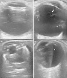
          Ocular B-mode ultrasonography (US) is an important adjuvant for the clinical assessment of a variety of ocular diseases. When ophthalmoscopy is not possible, mainly due to opacification of the transparent media (e.g., mature cataract or vitreous haemorrhage), US can guide the ophthalmologist in diagnosing disease and choosing treatment. The superficial location and cystic structure of the eye make US ideal for imaging of the eye. Moreover, dynamic study helps distinguish between various conditions that would otherwise be difficult to differentiate in some clinical setting, such as vitreous, retinal, and choroidal detachment. US is also good technique for detecting other pathologic conditions such as lens dislocation, vitreous haemorrhage, asteroid hyalosis, optic disc drusen, and tumors (e.g., choroidal melanoma, metastases, hemangioma). An understanding of the basic anatomy of the eye, the US technique, and common entities that affect the ocular globe will allow radiologists to offer this valuable imaging modality to patients and referring clinicians. This article focuses on the US anatomy and pathologic conditions that affect the posterior ocular segment.

          Teaching points

          • US is specially indicated when ocular fundus cannot be assessed on ophthalmoscopy.

          • Multipurpose equipment with high-frequency transducers is optimal for imaging the eye.

          • Ultrasound can reliably depict ocular anatomy and pathology as detachments and tumours.

          • Dynamic examination is vital for distinguishing certain pathologic conditions as detachments.

          Electronic supplementary material

          The online version of this article (doi:10.1007/s13244-016-0471-z) contains supplementary material, which is available to authorized users.

          Related collections

          Most cited references35

          • Record: found
          • Abstract: found
          • Article: not found

          High-resolution ultrasound imaging of the eye - a review.

          This report summarizes the physics, technology and clinical application of ultrasound biomicroscopy (UBM) of the eye, in which frequencies of 35 MHz and above provide over a threefold improvement in resolution compared with conventional ophthalmic ultrasound systems. UBM allows imaging of anatomy and pathology involving the anterior segment, including regions obscured by overlying optically opaque anatomic or pathologic structures. UBM provides diagnostically significant information in conditions such as glaucoma, cysts and neoplasms, trauma and foreign bodies. UBM also can provide crucial biometric information regarding anterior segment structures, including the cornea and its constituent layers and the anterior and posterior chambers. Although UBM has now been in use for over 15 years, new technologies, including transducer arrays, pulse encoding and combination of ultrasound with light, offer the potential for significant advances in high-resolution diagnostic imaging of the eye.
            Bookmark
            • Record: found
            • Abstract: found
            • Article: not found

            Imaging of orbital trauma.

            W S Kubal (2008)
            Assessing traumatic orbital injuries is an important challenge for radiologists; this assessment is even more difficult when the orbital injury is associated with injuries involving multiple organs. Common posttraumatic orbital injuries include anterior chamber injuries, injuries to the lens, open-globe injuries, ocular detachments, intraorbital foreign bodies, carotid cavernous fistula, and optic nerve injuries. Radiographic examination of the orbits is rarely performed. Ultrasonography (US) can be very useful for evaluating the globe and its contents; however, US is contraindicated if a ruptured globe is suspected. Magnetic resonance imaging may be difficult to perform emergently; it is contraindicated if there is a possibility that a metallic intraorbital foreign body is present. Computed tomography (CT) is considered to be the top choice for evaluating orbital trauma. The best protocol is to obtain thin-section axial CT scans, then to perform multiplanar reformation. When evaluating a patient with an orbital injury, the radiologist should do the following: (a) evaluate the bony orbit for fractures, note any herniations of orbital contents, and pay particular attention to the orbital apex; (b) evaluate the anterior chamber; (c) evaluate the position of the lens (the lens may be displaced, and it may be either completely or partially dislocated); (d) evaluate the posterior segment of the globe, look for bleeds or abnormal fluid collections, and evaluate for radiopaque or radiolucent foreign bodies; and (e) evaluate the ophthalmic veins and the optic nerve complex, especially the orbital apex. (c) RSNA, 2008.
              Bookmark
              • Record: found
              • Abstract: found
              • Article: found
              Is Open Access

              Strategies for improving early detection and diagnosis of neovascular age-related macular degeneration

              Treatment of the neovascular form of age-related macular degeneration (AMD) has been revolutionized by the introduction of such agents as ranibizumab, bevacizumab, and aflibercept. As a result, the incidence of legal blindness occurring secondary to AMD has fallen dramatically in recent years in many countries. While these agents have undoubtedly been successful in reducing visual impairment and blindness, patients with neovascular AMD typically lose some vision over time, and often lose the ability to read, drive, or perform other important activities of daily living. Efforts are therefore under way to develop strategies that allow for earlier detection and treatment of this disease. In this review, we begin by providing an overview of the rationale for, and the benefits of, early detection and treatment of neovascular AMD. To achieve this, we begin by providing an overview of the pathophysiology and natural history of choroidal neovascularization, before reviewing the evidence from both clinical trials and “real-world” outcome studies. We continue by highlighting an area that is often overlooked: the importance of patient education and awareness for early AMD detection. We conclude the review by reviewing an array of both established and emerging technologies for early detection of choroidal neovascularization, ranging from Amsler chart testing, to hyperacuity testing, to advanced imaging techniques, such as optical coherence tomography.
                Bookmark

                Author and article information

                Contributors
                0034 627 229 631 , mdelahozpolo@gmail.com
                Journal
                Insights Imaging
                Insights Imaging
                Insights into Imaging
                Springer Berlin Heidelberg (Berlin/Heidelberg )
                1869-4101
                24 February 2016
                24 February 2016
                June 2016
                : 7
                : 3
                : 351-364
                Affiliations
                [ ]Radiology Department, Hospital Sant Pau i Santa Tecla, Rambla Vella 14, 43003 Tarragona, Spain
                [ ]Hospital El Vendrell, Carretera de Barcelona, s/n, 43700 El Vendrell, Spain
                [ ]Radiology Department, Hospital de Viladecans, Viladecans, Spain
                [ ]Ophthalmology Department, Hospital de Viladecans, Viladecans, Spain
                [ ]Ophthalmology Department, Hospital Universitari de Bellvitge, L’Hospitalet de Llobregat, Spain
                Article
                471
                10.1007/s13244-016-0471-z
                4877345
                26910565
                adb08acd-40d9-43e7-9e06-f94534c6e385
                © The Author(s) 2016

                Open Access This article is distributed under the terms of the Creative Commons Attribution 4.0 International License (http://creativecommons.org/licenses/by/4.0/), which permits unrestricted use, distribution, and reproduction in any medium, provided you give appropriate credit to the original author(s) and the source, provide a link to the Creative Commons license, and indicate if changes were made.

                History
                : 4 November 2015
                : 23 January 2016
                : 26 January 2016
                Categories
                Pictorial Review
                Custom metadata
                © The Author(s) 2016

                Radiology & Imaging
                ultrasound,posterior eye segment,retina,vitreous body,choroid
                Radiology & Imaging
                ultrasound, posterior eye segment, retina, vitreous body, choroid

                Comments

                Comment on this article