3
views
0
recommends
+1 Recommend
0 collections
    0
    shares
      • Record: found
      • Abstract: found
      • Article: found
      Is Open Access

      Magnetic resonance imaging of the orbit, Part 2: Characterization of orbital pathologies

      review-article

      Read this article at

      Bookmark
          There is no author summary for this article yet. Authors can add summaries to their articles on ScienceOpen to make them more accessible to a non-specialist audience.

          Abstract

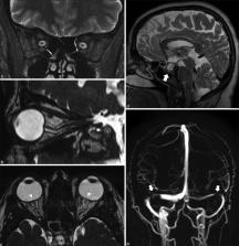
          In this article we focus on a systematic approach to assess common orbital lesions on magnetic resonance imaging (MRI). The identification of the probable compartment or structure of origin helps narrow the differential diagnosis of a lesion. Analyzing the morphology, appearance, and signal intensity on various sequences, the pattern, and degree of contrast enhancement are key to characterize lesions on MRI. Imaging features suggesting cellularity and vascularity can also be determined to help plan for biopsy or surgery of these lesions. MRI can also distinguish active from chronic disease in certain pathologies and aids in selecting appropriate medical management. MRI may thus serve as a diagnostic tool and help in guiding therapeutic strategies and posttreatment follow-up.

          Related collections

          Most cited references62

          • Record: found
          • Abstract: found
          • Article: found

          The 2016 World Health Organization Classification of Tumors of the Central Nervous System: a summary.

          The 2016 World Health Organization Classification of Tumors of the Central Nervous System is both a conceptual and practical advance over its 2007 predecessor. For the first time, the WHO classification of CNS tumors uses molecular parameters in addition to histology to define many tumor entities, thus formulating a concept for how CNS tumor diagnoses should be structured in the molecular era. As such, the 2016 CNS WHO presents major restructuring of the diffuse gliomas, medulloblastomas and other embryonal tumors, and incorporates new entities that are defined by both histology and molecular features, including glioblastoma, IDH-wildtype and glioblastoma, IDH-mutant; diffuse midline glioma, H3 K27M-mutant; RELA fusion-positive ependymoma; medulloblastoma, WNT-activated and medulloblastoma, SHH-activated; and embryonal tumour with multilayered rosettes, C19MC-altered. The 2016 edition has added newly recognized neoplasms, and has deleted some entities, variants and patterns that no longer have diagnostic and/or biological relevance. Other notable changes include the addition of brain invasion as a criterion for atypical meningioma and the introduction of a soft tissue-type grading system for the now combined entity of solitary fibrous tumor / hemangiopericytoma-a departure from the manner by which other CNS tumors are graded. Overall, it is hoped that the 2016 CNS WHO will facilitate clinical, experimental and epidemiological studies that will lead to improvements in the lives of patients with brain tumors.
            Bookmark
            • Record: found
            • Abstract: found
            • Article: not found

            Soft-tissue tumors and tumorlike lesions: a systematic imaging approach.

            Soft-tissue lesions are frequently encountered by radiologists in everyday clinical practice. Characterization of these soft-tissue lesions remains problematic, despite advances in imaging. By systematically using clinical history, lesion location, mineralization on radiographs, and signal intensity characteristics on magnetic resonance images, one can (a) determine the diagnosis for the subset of determinate lesions that have characteristic clinical and imaging features and (b) narrow the differential diagnosis for lesions that demonstrate indeterminate characteristics. If a lesion cannot be characterized as a benign entity, the lesion should be reported as indeterminate, and the patient should undergo biopsy to exclude malignancy. (c) RSNA, 2009.
              Bookmark
              • Record: found
              • Abstract: not found
              • Article: not found

              Imaging findings of fibrous dysplasia with histopathologic and intraoperative correlation.

                Bookmark

                Author and article information

                Journal
                Indian J Ophthalmol
                Indian J Ophthalmol
                IJO
                Indian J Ophthalmol
                Indian Journal of Ophthalmology
                Wolters Kluwer - Medknow (India )
                0301-4738
                1998-3689
                October 2021
                25 September 2021
                : 69
                : 10
                : 2585-2616
                Affiliations
                [1]Neurovascular and Interventional Radiology, Apollo Speciality Hospital, Bengaluru, Karnataka, India
                [1 ]Orbit & Oculoplasty, Narayana Nethralaya, Bengaluru, Karnataka, India
                [2 ]Division of Neuroradiology, Department of Medical Imaging, The Ottawa Hospital - Civic Campus, Ottawa, Canada
                [3 ]Orbit & Oculoplasty, Centre for Sight, Hyderabad, Telangana, India
                Author notes
                Correspondence to: Dr. Chinmay Nagesh, Consultant, Neurovascular & Interventional Radiology, Apollo Speciality Hospital, #2, 14th Cross, 3 rd Block, Jayanagar, Bengaluru - 560 011, Karnataka, India. E-mail: drchinmaynagesh@ 123456gmail.com
                Article
                IJO-69-2585
                10.4103/ijo.IJO_904_21
                8597442
                34571598
                d6a7d66b-50c9-4ff6-9a68-dd4764f264dc
                Copyright: © 2021 Indian Journal of Ophthalmology

                This is an open-access article distributed under the terms of the Creative Commons Attribution-Noncommercial-Share Alike 4.0 Unported, which permits unrestricted use, distribution, and reproduction in any medium, provided the original work is properly cited.

                History
                : 17 April 2021
                : 07 July 2021
                : 12 August 2021
                Categories
                Review Article

                Ophthalmology & Optometry
                magnetic resonance imaging,mri,orbit,pathology,tumor
                Ophthalmology & Optometry
                magnetic resonance imaging, mri, orbit, pathology, tumor

                Comments

                Comment on this article