25
views
0
recommends
+1 Recommend
0 collections
    0
    shares
      • Record: found
      • Abstract: found
      • Article: found
      Is Open Access

      CCM2–CCM3 interaction stabilizes their protein expression and permits endothelial network formation

      research-article

      Read this article at

      Bookmark
          There is no author summary for this article yet. Authors can add summaries to their articles on ScienceOpen to make them more accessible to a non-specialist audience.

          Abstract

          CCM2–CCM3 interactions protect CCM2 and CCM3 from proteasomal degradation, and both CCM2 and CCM3 are required for normal endothelial cell network formation.

          Abstract

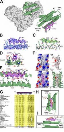
          Mutations in the essential adaptor proteins CCM2 or CCM3 lead to cerebral cavernous malformations (CCM), vascular lesions that most frequently occur in the brain and are strongly associated with hemorrhagic stroke, seizures, and other neurological disorders. CCM2 binds CCM3, but the molecular basis of this interaction, and its functional significance, have not been elucidated. Here, we used x-ray crystallography and structure-guided mutagenesis to show that an α-helical LD-like motif within CCM2 binds the highly conserved “HP1” pocket of the CCM3 focal adhesion targeting (FAT) homology domain. By knocking down CCM2 or CCM3 and rescuing with binding-deficient mutants, we establish that CCM2–CCM3 interactions protect CCM2 and CCM3 proteins from proteasomal degradation and show that both CCM2 and CCM3 are required for normal endothelial cell network formation. However, CCM3 expression in the absence of CCM2 is sufficient to support normal cell growth, revealing complex-independent roles for CCM3.

          Related collections

          Most cited references25

          • Record: found
          • Abstract: found
          • Article: not found

          ALINE: a WYSIWYG protein-sequence alignment editor for publication-quality alignments.

          Marked-up sequence alignments typically provide the central figure in articles describing proteins, whether in the fields of biochemistry, bioinformatics or structural biology. The generation of these figures is often unwieldy: interactive programs are often aesthetically limited and the use of batch programs requires the repetitive iterative editing of scripts. ALINE is a portable interactive graphical sequence-alignment editor implemented in Perl/Tk which produces publication-quality sequence-alignment figures where "what you see is what you get". ALINE is freely available for download from http://crystal.bcs.uwa.edu.au/px/charlie/software/aline/.
            Bookmark
            • Record: found
            • Abstract: found
            • Article: not found

            The molecular basis of filamin binding to integrins and competition with talin.

            The ability of adhesion receptors to transmit biochemical signals and mechanical force across cell membranes depends on interactions with the actin cytoskeleton. Filamins are large, actin-crosslinking proteins that connect multiple transmembrane and signaling proteins to the cytoskeleton. Here, we describe the high-resolution structure of an interface between filamin A and an integrin adhesion receptor. When bound, the integrin beta cytoplasmic tail forms an extended beta strand that interacts with beta strands C and D of the filamin immunoglobulin-like domain (IgFLN) 21. This interface is common to many integrins, and we suggest it is a prototype for other IgFLN domain interactions. Notably, the structurally defined filamin binding site overlaps with that of the integrin-regulator talin, and these proteins compete for binding to integrin tails, allowing integrin-filamin interactions to impact talin-dependent integrin activation. Phosphothreonine-mimicking mutations inhibit filamin, but not talin, binding, indicating that kinases may modulate this competition and provide additional means to control integrin functions.
              Bookmark
              • Record: found
              • Abstract: found
              • Article: not found

              Mutations in a gene encoding a novel protein containing a phosphotyrosine-binding domain cause type 2 cerebral cavernous malformations.

              Cerebral cavernous malformations (CCMs) are congenital vascular anomalies of the central nervous system that can result in hemorrhagic stroke, seizures, recurrent headaches, and focal neurologic deficits. Mutations in the gene KRIT1 are responsible for type 1 CCM (CCM1). We report that a novel gene, MGC4607, exhibits eight different mutations in nine families with type 2 CCM (CCM2). MGC4607, similar to the KRIT1 binding partner ICAP1alpha, encodes a protein with a phosphotyrosine-binding domain. This protein may be part of the complex pathway of integrin signaling that, when perturbed, causes abnormal vascular morphogenesis in the brain, leading to CCM formation.
                Bookmark

                Author and article information

                Journal
                J Cell Biol
                J. Cell Biol
                jcb
                jcb
                The Journal of Cell Biology
                The Rockefeller University Press
                0021-9525
                1540-8140
                30 March 2015
                : 208
                : 7
                : 987-1001
                Affiliations
                [1 ]Department of Pharmacology and [2 ]Department of Cell Biology, Yale University, New Haven, CT 06520
                Author notes
                Correspondence to Titus J. Boggon: titus.boggon@ 123456yale.edu ; or David A. Calderwood: david.calderwood@ 123456yale.edu
                [*]

                K.M. Draheim and X. Li contributed equally to this paper.

                Article
                201407129
                10.1083/jcb.201407129
                4384732
                25825518
                ab4329b4-d73a-4f36-bd39-111ba035066f
                © 2015 Draheim et al.

                This article is distributed under the terms of an Attribution–Noncommercial–Share Alike–No Mirror Sites license for the first six months after the publication date (see http://www.rupress.org/terms). After six months it is available under a Creative Commons License (Attribution–Noncommercial–Share Alike 3.0 Unported license, as described at http://creativecommons.org/licenses/by-nc-sa/3.0/).

                History
                : 28 July 2014
                : 19 February 2015
                Categories
                Research Articles
                Article

                Cell biology
                Cell biology

                Comments

                Comment on this article