6
views
0
recommends
+1 Recommend
0 collections
    0
    shares
      • Record: found
      • Abstract: found
      • Article: found
      Is Open Access

      Recent advances in cerebral cavernous malformation research

      research-article

      Read this article at

      Bookmark
          There is no author summary for this article yet. Authors can add summaries to their articles on ScienceOpen to make them more accessible to a non-specialist audience.

          Abstract

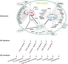
          Cerebral cavernous malformations (CCM) are manifested by microvascular lesions characterized by leaky endothelial cells with minimal intervening parenchyma predominantly in the central nervous system predisposed to hemorrhagic stroke, resulting in focal neurological defects. Till date, three proteins are implicated in this condition: CCM1 (KRIT1), CCM2 (MGC4607), and CCM3 (PDCD10). These multi-domain proteins form a protein complex via CCM2 that function as a docking site for the CCM signaling complex, which modulates many signaling pathways. Defects in the formation of this signaling complex have been shown to affect a wide range of cellular processes including cell-cell contact stability, vascular angiogenesis, oxidative damage protection and multiple biogenic events. In this review we provide an update on recent advances in structure and function of these CCM proteins, especially focusing on the signaling cascades involved in CCM pathogenesis and the resultant CCM cellular phenotypes in the past decade.

          Related collections

          Most cited references156

          • Record: found
          • Abstract: found
          • Article: not found

          The Nrf2 cell defence pathway: Keap1-dependent and -independent mechanisms of regulation.

          The transcription factor Nrf2 (NF-E2-related factor 2) plays a vital role in maintaining cellular homeostasis, especially upon the exposure of cells to chemical or oxidative stress, through its ability to regulate the basal and inducible expression of a multitude of antioxidant proteins, detoxification enzymes and xenobiotic transporters. In addition, Nrf2 contributes to diverse cellular functions including differentiation, proliferation, inflammation and lipid synthesis and there is an increasing association of aberrant expression and/or function of Nrf2 with pathologies including cancer, neurodegeneration and cardiovascular disease. The activity of Nrf2 is primarily regulated via its interaction with Keap1 (Kelch-like ECH-associated protein 1), which directs the transcription factor for proteasomal degradation. Although it is generally accepted that modification (e.g. chemical adduction, oxidation, nitrosylation or glutathionylation) of one or more critical cysteine residues in Keap1 represents a likely chemico-biological trigger for the activation of Nrf2, unequivocal evidence for such a phenomenon remains elusive. An increasing body of literature has revealed alternative mechanisms of Nrf2 regulation, including phosphorylation of Nrf2 by various protein kinases (PKC, PI3K/Akt, GSK-3β, JNK), interaction with other protein partners (p21, caveolin-1) and epigenetic factors (micro-RNAs -144, -28 and -200a, and promoter methylation). These and other processes are potentially important determinants of Nrf2 activity, and therefore may contribute to the maintenance of cellular homeostasis. Here, we dissect evidence supporting these Keap1-dependent and -independent mechanisms of Nrf2 regulation. Furthermore, we highlight key knowledge gaps in this important field of biology, and suggest how these may be addressed experimentally. Copyright © 2012 Elsevier Inc. All rights reserved.
            Bookmark
            • Record: found
            • Abstract: found
            • Article: found
            Is Open Access

            Antioxidant responses and cellular adjustments to oxidative stress

            Redox biological reactions are now accepted to bear the Janus faceted feature of promoting both physiological signaling responses and pathophysiological cues. Endogenous antioxidant molecules participate in both scenarios. This review focuses on the role of crucial cellular nucleophiles, such as glutathione, and their capacity to interact with oxidants and to establish networks with other critical enzymes such as peroxiredoxins. We discuss the importance of the Nrf2-Keap1 pathway as an example of a transcriptional antioxidant response and we summarize transcriptional routes related to redox activation. As examples of pathophysiological cellular and tissular settings where antioxidant responses are major players we highlight endoplasmic reticulum stress and ischemia reperfusion. Topologically confined redox-mediated post-translational modifications of thiols are considered important molecular mechanisms mediating many antioxidant responses, whereas redox-sensitive microRNAs have emerged as key players in the posttranscriptional regulation of redox-mediated gene expression. Understanding such mechanisms may provide the basis for antioxidant-based therapeutic interventions in redox-related diseases.
              Bookmark
              • Record: found
              • Abstract: found
              • Article: not found

              The small GTP-binding protein rho regulates the assembly of focal adhesions and actin stress fibers in response to growth factors.

              Actin stress fibers are one of the major cytoskeletal structures in fibroblasts and are linked to the plasma membrane at focal adhesions. rho, a ras-related GTP-binding protein, rapidly stimulated stress fiber and focal adhesion formation when microinjected into serum-starved Swiss 3T3 cells. Readdition of serum produced a similar response, detectable within 2 min. This activity was due to a lysophospholipid, most likely lysophosphatidic acid, bound to serum albumin. Other growth factors including PDGF induced actin reorganization initially to form membrane ruffles, and later, after 5 to 10 min, stress fibers. For all growth factors tested the stimulation of focal adhesion and stress fiber assembly was inhibited when endogenous rho function was blocked, whereas membrane ruffling was unaffected. These data imply that rho is essential specifically for the coordinated assembly of focal adhesions and stress fibers induced by growth factors.
                Bookmark

                Author and article information

                Journal
                101726367
                47542
                Vessel Plus
                Vessel Plus
                Vessel plus
                2574-1209
                24 July 2019
                28 August 2018
                2018
                29 July 2019
                : 2
                : 21
                Affiliations
                Department of Biomedical Sciences, Texas Tech University Health Science Center El Paso, El Paso, TX 79905, USA
                Author notes

                Author’s contributions Akhil Padarti drafted the manuscript under the supervision of Jun Zhang.

                Correspondence to: Dr. Jun Zhang, Department of Biomedical Sciences, Texas Tech Udniversity Health Science Center, 5001 El Paso Drive, El Paso, El Paso, TX 79905, USA. jun.zhang2000@ 123456gmail.com
                Article
                NIHMS1043190
                10.20517/2574-1209.2018.34
                6663315
                31360916
                3aa2c2d0-2362-4da2-960d-c2edb8ad7bfd

                Open Access This article is licensed under a Creative Commons Attribution 4.0 International License ( https://creativecommons.org/licenses/by/4.0/), which permits unrestricted use, sharing, adaptation, distribution and reproduction in any medium or format, for any purpose, even commercially, as long as you give appropriate credit to the original author(s) and the source, provide a link to the Creative Commons license, and indicate if changes were made.

                History
                Categories
                Article

                cerebral cavernous malformation,cerebral cavernous malformation signaling complex,angiogenesis,endothelial cells,cellular function,microvessel lesions,protein structure,function domain,motif

                Comments

                Comment on this article