157
views
0
recommends
+1 Recommend
0 collections
    0
    shares
      • Record: found
      • Abstract: found
      • Article: found
      Is Open Access

      Antioxidant responses and cellular adjustments to oxidative stress

      review-article

      Read this article at

      Bookmark
          There is no author summary for this article yet. Authors can add summaries to their articles on ScienceOpen to make them more accessible to a non-specialist audience.

          Abstract

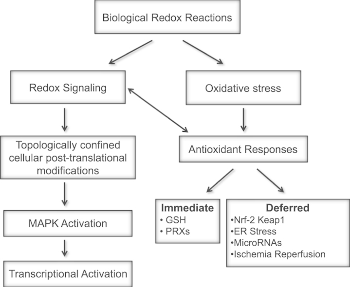
          Redox biological reactions are now accepted to bear the Janus faceted feature of promoting both physiological signaling responses and pathophysiological cues. Endogenous antioxidant molecules participate in both scenarios. This review focuses on the role of crucial cellular nucleophiles, such as glutathione, and their capacity to interact with oxidants and to establish networks with other critical enzymes such as peroxiredoxins. We discuss the importance of the Nrf2-Keap1 pathway as an example of a transcriptional antioxidant response and we summarize transcriptional routes related to redox activation. As examples of pathophysiological cellular and tissular settings where antioxidant responses are major players we highlight endoplasmic reticulum stress and ischemia reperfusion. Topologically confined redox-mediated post-translational modifications of thiols are considered important molecular mechanisms mediating many antioxidant responses, whereas redox-sensitive microRNAs have emerged as key players in the posttranscriptional regulation of redox-mediated gene expression. Understanding such mechanisms may provide the basis for antioxidant-based therapeutic interventions in redox-related diseases.

          Graphical abstract

          Highlights

          • Antioxidant responses are crucial for both redox signaling and redox damage.

          • Glutathione-mediated reactions and Nrf2-Keap1 pathway are key antioxidant responses.

          • Redox-related post-translational modifications activate specific signaling pathways.

          • Redox-sensitive microRNAs contribute to redox-mediated gene expression regulation.

          • ER stress and ischemia-reperfusion are antioxidant-related pathophysiological events.

          Related collections

          Most cited references180

          • Record: found
          • Abstract: found
          • Article: not found

          Posttranscriptional regulation of the heterochronic gene lin-14 by lin-4 mediates temporal pattern formation in C. elegans.

          During C. elegans development, the temporal pattern of many cell lineages is specified by graded activity of the heterochronic gene Lin-14. Here we demonstrate that a temporal gradient in Lin-14 protein is generated posttranscriptionally by multiple elements in the lin-14 3'UTR that are regulated by the heterochronic gene Lin-4. The lin-14 3'UTR is both necessary and sufficient to confer lin-4-mediated posttranscriptional temporal regulation. The function of the lin-14 3'UTR is conserved between C. elegans and C. briggsae. Among the conserved sequences are seven elements that are each complementary to the lin-4 RNAs. A reporter gene bearing three of these elements shows partial temporal gradient activity. These data suggest a molecular mechanism for Lin-14p temporal gradient formation: the lin-4 RNAs base pair to sites in the lin-14 3'UTR to form multiple RNA duplexes that down-regulate lin-14 translation.
            Bookmark
            • Record: found
            • Abstract: found
            • Article: not found

            Physiological and pathological roles for microRNAs in the immune system.

            Mammalian microRNAs (miRNAs) have recently been identified as important regulators of gene expression, and they function by repressing specific target genes at the post-transcriptional level. Now, studies of miRNAs are resolving some unsolved issues in immunology. Recent studies have shown that miRNAs have unique expression profiles in cells of the innate and adaptive immune systems and have pivotal roles in the regulation of both cell development and function. Furthermore, when miRNAs are aberrantly expressed they can contribute to pathological conditions involving the immune system, such as cancer and autoimmunity; they have also been shown to be useful as diagnostic and prognostic indicators of disease type and severity. This Review discusses recent advances in our understanding of both the intended functions of miRNAs in managing immune cell biology and their pathological roles when their expression is dysregulated.
              Bookmark
              • Record: found
              • Abstract: found
              • Article: not found

              Reactive oxygen species promote TNFalpha-induced death and sustained JNK activation by inhibiting MAP kinase phosphatases.

              TNFalpha is a pleiotropic cytokine that induces either cell proliferation or cell death. Inhibition of NF-kappaB activation increases susceptibility to TNFalpha-induced death, concurrent with sustained JNK activation, an important contributor to the death response. Sustained JNK activation in NF-kappaB-deficient cells was suggested to depend on reactive oxygen species (ROS), but how ROS affect JNK activation was unclear. We now show that TNFalpha-induced ROS, whose accumulation is suppressed by mitochondrial superoxide dismutase, cause oxidation and inhibition of JNK-inactivating phosphatases by converting their catalytic cysteine to sulfenic acid. This results in sustained JNK activation, which is required for cytochrome c release and caspase 3 cleavage, as well as necrotic cell death. Treatment of cells or experimental animals with an antioxidant prevents H(2)O(2) accumulation, JNK phosphatase oxidation, sustained JNK activity, and both forms of cell death. Antioxidant treatment also prevents TNFalpha-mediated fulminant liver failure without affecting liver regeneration.
                Bookmark

                Author and article information

                Contributors
                Journal
                Redox Biol
                Redox Biol
                Redox Biology
                Elsevier
                2213-2317
                21 July 2015
                December 2015
                21 July 2015
                : 6
                : 183-197
                Affiliations
                [a ]Department of Cell Biology and Immunology, Centro de Biología Molecular “Severo Ochoa” (CSIC-UAM), Nicolás Cabrera 1, 28049 Madrid, Spain
                [b ]Faculty of Biochemistry and Molecular Medicine, Biocenter Oulu, Aapistie 7, University of Oulu, FI-90230 Oulu, Finland
                [c ]Centro de Biología Molecular “Severo Ochoa” (CSIC-UAM) and Departamento de Biología Molecular, Universidad Autónoma de Madrid, Nicolás Cabrera 1, 28049 Madrid, Spain; Instituto de Investigación Sanitaria Princesa (IIS-IP), 28006 Madrid, Spain
                Author notes
                [* ]Corresponding author. slamas@ 123456cbm.csic.es
                Article
                S2213-2317(15)00080-4
                10.1016/j.redox.2015.07.008
                4534574
                26233704
                9239d0a3-287b-4d3f-b7f3-48fd3662ebcd
                © 2015 Published by Elsevier B.V.

                This is an open access article under the CC BY-NC-ND license (http://creativecommons.org/licenses/by-nc-nd/4.0/).

                History
                : 30 June 2015
                : 6 July 2015
                Categories
                Review Article

                redox signaling,antioxidants,transcription factors,ischemia–reperfusion,er stress

                Comments

                Comment on this article