10
views
0
recommends
+1 Recommend
0 collections
    0
    shares
      • Record: found
      • Abstract: found
      • Article: found
      Is Open Access

      Cerebrospinal fluid tracer efflux to parasagittal dura in humans

      research-article
      1 , 2 , 3 ,
      Nature Communications
      Nature Publishing Group UK
      Neuroscience, Neurological disorders

      Read this article at

      Bookmark
          There is no author summary for this article yet. Authors can add summaries to their articles on ScienceOpen to make them more accessible to a non-specialist audience.

          Abstract

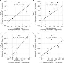
          The mechanisms behind molecular transport from cerebrospinal fluid to dural lymphatic vessels remain unknown. This study utilized magnetic resonance imaging along with cerebrospinal fluid tracer to visualize clearance pathways to human dural lymphatics in vivo. In 18 subjects with suspicion of various types of cerebrospinal fluid disorders, 3D T2-Fluid Attenuated Inversion Recovery, T1-black-blood, and T1 gradient echo acquisitions were obtained prior to intrathecal administration of the contrast agent gadobutrol (0.5 ml, 1 mmol/ml), serving as a cerebrospinal fluid tracer. Propagation of tracer was followed with T1 sequences at 3, 6, 24 and 48 h after the injection. The tracer escaped from cerebrospinal fluid into parasagittal dura along the superior sagittal sinus at areas nearby entry of cortical cerebral veins. The findings demonstrate that trans-arachnoid molecular passage does occur and suggest that parasagittal dura may serve as a bridging link between human brain and dural lymphatic vessels.

          Abstract

          Mechanisms behind molecular transport from cerebrospinal fluid to dural lymphatic vessels remain unknown. This study demonstrates that trans-arachnoid molecular passage does occur and suggests that parasagittal dura may serve as a bridging link between human brain and dural lymphatic vessels.

          Related collections

          Most cited references23

          • Record: found
          • Abstract: found
          • Article: not found

          A paravascular pathway facilitates CSF flow through the brain parenchyma and the clearance of interstitial solutes, including amyloid β.

          Because it lacks a lymphatic circulation, the brain must clear extracellular proteins by an alternative mechanism. The cerebrospinal fluid (CSF) functions as a sink for brain extracellular solutes, but it is not clear how solutes from the brain interstitium move from the parenchyma to the CSF. We demonstrate that a substantial portion of subarachnoid CSF cycles through the brain interstitial space. On the basis of in vivo two-photon imaging of small fluorescent tracers, we showed that CSF enters the parenchyma along paravascular spaces that surround penetrating arteries and that brain interstitial fluid is cleared along paravenous drainage pathways. Animals lacking the water channel aquaporin-4 (AQP4) in astrocytes exhibit slowed CSF influx through this system and a ~70% reduction in interstitial solute clearance, suggesting that the bulk fluid flow between these anatomical influx and efflux routes is supported by astrocytic water transport. Fluorescent-tagged amyloid β, a peptide thought to be pathogenic in Alzheimer's disease, was transported along this route, and deletion of the Aqp4 gene suppressed the clearance of soluble amyloid β, suggesting that this pathway may remove amyloid β from the central nervous system. Clearance through paravenous flow may also regulate extracellular levels of proteins involved with neurodegenerative conditions, its impairment perhaps contributing to the mis-accumulation of soluble proteins.
            Bookmark
            • Record: found
            • Abstract: found
            • Article: not found

            Structural and functional features of central nervous system lymphatics

            One of the characteristics of the CNS is the lack of a classical lymphatic drainage system. Although it is now accepted that the CNS undergoes constant immune surveillance that takes place within the meningeal compartment 1–3 , the mechanisms governing the entrance and exit of immune cells from the CNS remain poorly understood 4–6 . In searching for T cell gateways into and out of the meninges, we discovered functional lymphatic vessels lining the dural sinuses. These structures express all of the molecular hallmarks of lymphatic endothelial cells, are able to carry both fluid and immune cells from the CSF, and are connected to the deep cervical lymph nodes. The unique location of these vessels may have impeded their discovery to date, thereby contributing to the long-held concept of the absence of lymphatic vasculature in the CNS. The discovery of the CNS lymphatic system may call for a reassessment of basic assumptions in neuroimmunology and shed new light on the etiology of neuroinflammatory and neurodegenerative diseases associated with immune system dysfunction.
              Bookmark
              • Record: found
              • Abstract: found
              • Article: found
              Is Open Access

              A dural lymphatic vascular system that drains brain interstitial fluid and macromolecules

              Aspelund et al. discover the presence of a lymphatic vessel network in the dura mater of the mouse brain and show that these dural lymphatic vessels are important for the clearance of macromolecules from the brain.
                Bookmark

                Author and article information

                Contributors
                p.k.eide@medisin.uio.no
                Journal
                Nat Commun
                Nat Commun
                Nature Communications
                Nature Publishing Group UK (London )
                2041-1723
                17 January 2020
                17 January 2020
                2020
                : 11
                : 354
                Affiliations
                [1 ]ISNI 0000 0004 0389 8485, GRID grid.55325.34, Department of Radiology and Nuclear Medicine, , Oslo University Hospital - Rikshospitalet, ; Oslo, Norway
                [2 ]ISNI 0000 0004 0389 8485, GRID grid.55325.34, Department of Neurosurgery, , Oslo University Hospital-Rikshospitalet, ; Oslo, Norway
                [3 ]ISNI 0000 0004 1936 8921, GRID grid.5510.1, Institute of Clinical Medicine, Faculty of Medicine, , University of Oslo, ; Oslo, Norway
                Author information
                http://orcid.org/0000-0003-0919-4510
                http://orcid.org/0000-0001-6881-9280
                Article
                14195
                10.1038/s41467-019-14195-x
                6969040
                31953399
                ab0d7099-a448-4fb6-b948-20c36654e932
                © The Author(s) 2020

                Open Access This article is licensed under a Creative Commons Attribution 4.0 International License, which permits use, sharing, adaptation, distribution and reproduction in any medium or format, as long as you give appropriate credit to the original author(s) and the source, provide a link to the Creative Commons license, and indicate if changes were made. The images or other third party material in this article are included in the article’s Creative Commons license, unless indicated otherwise in a credit line to the material. If material is not included in the article’s Creative Commons license and your intended use is not permitted by statutory regulation or exceeds the permitted use, you will need to obtain permission directly from the copyright holder. To view a copy of this license, visit http://creativecommons.org/licenses/by/4.0/.

                History
                : 17 June 2019
                : 17 December 2019
                Categories
                Article
                Custom metadata
                © The Author(s) 2020

                Uncategorized
                neuroscience,neurological disorders
                Uncategorized
                neuroscience, neurological disorders

                Comments

                Comment on this article