8
views
0
recommends
+1 Recommend
0 collections
    0
    shares
      • Record: found
      • Abstract: found
      • Article: found
      Is Open Access

      Evaluating glaucomatous abnormality in peripapillary optical coherence tomography enface visualisation of the retinal nerve fibre layer reflectance

      research-article

      Read this article at

      Bookmark
          There is no author summary for this article yet. Authors can add summaries to their articles on ScienceOpen to make them more accessible to a non-specialist audience.

          Abstract

          Purpose

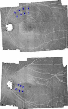
          Optical coherence tomography ( OCT) enface visualisation of the retinal nerve fibre layer ( RNFL) reflectance has been found to have some advantages over retinal thickness measures. However, it is not yet clear how abnormalities on enface images relate to findings of abnormalities from other clinical measures such as the circumpapillary retinal nerve fibre layer thickness ( cRNFLT). We developed a technique to analyse the RNFL reflectance on the OCT enface images, and to investigate its relation with the cRNFLT.

          Methods

          Spectralis ( http://www.heidelbergengineering.com) OCT scans of the central retinal ±24° were analysed in the study eye of 31 controls and 33 patients, ages 61 (±9) and 69 (±8) years respectively. Enface slab‐images were extracted at 16–24, 24–36, and 24–52 μm from the inner limiting membrane in the temporal raphe, perifoveal and disc regions respectively. Reflectance probability maps were generated for the patients based on the control data. Glaucomatous abnormality was defined on the slab‐images when the slab‐area with reflectance abnormality was greater than the 95th percentile, and on the cRNFLT when the thickness measure was less than the fifth percentile, of that found in controls. The fraction of slab‐image showing reflectance abnormality was compared to cRNFLT in the patient group, using Spearman's rho. Agreement between the findings of abnormality based on cRNFLT and slab‐image reflectance was assessed using Cohen's kappa.

          Results

          Slab‐image and cRNFLT findings were in agreement for 26/33 eyes; four subjects showed cRNFLT abnormality but not slab‐image abnormality, and three subjects showed slab‐image abnormality but not cRNFLT abnormality. Spearman's rho found r s(31) = −0.82. The reflectance findings and cRNFLT findings were consistent in 27/33 for both the superior temporal ( ST) and inferior temporal ( IT) sectors, and Cohen's kappa found 0.53 and 0.61 respectively.

          Conclusion

          The surface area of enface slab‐images showing RNFL reflectance were strongly related to the cRNFLT measures, and the classification of a subject with glaucoma based on enface reflectance findings and cRNFLT findings had a generally good agreement. The larger retinal area assessed by the enface method preserves the spatial location of the RNFL abnormalities, and makes the technique a useful approach for identifying regions of potential RNFL abnormality for targeted perimetry.

          Related collections

          Most cited references23

          • Record: found
          • Abstract: found
          • Article: not found

          Morphologic changes in the lamina cribrosa correlated with neural loss in open-angle glaucoma.

          We divided 25 glaucomatous human eyes into three groups representing mild (Group 1, seven eyes), moderate (Group 2, 11 eyes), and severe (Group 3, six eyes) optic nerve damage, based on visual field testing or remaining number of optic nerve fibers. The optic nerve head of each eye was examined by scanning electron microscopy. Compression of the successive lamina cribrosa sheets was the earliest detected abnormality, occurring in some eyes before the detection of visual field loss. Backward bowing of the entire lamina cribrosa was a later change and involved its upper and lower poles more than the mid-nerve head. The diameter of the scleral opening at the level of Bruch's membrane did not enlarge in these adult glaucomatous eyes. Mechanical compression of the nerve head occurred early enough to be considered a primary pathogenetic event in glaucomatous damage.
            Bookmark
            • Record: found
            • Abstract: found
            • Article: not found

            Shadow removal and contrast enhancement in optical coherence tomography images of the human optic nerve head.

            To improve the quality of optical coherence tomography (OCT) images of the optic nerve head (ONH). Two algorithms were developed, one to compensate for light attenuation and the other to enhance contrast in OCT images. The former was borrowed from developments in ultrasound imaging and was proven suitable with either time- or spectral-domain OCT. The latter was based on direct application of pixel intensity exponentiation. The performances of these two algorithms were tested on spectral-domain OCT images of four adult ONHs. Application of the compensation algorithm significantly reduced the intralayer contrast (from 0.74 ± 0.16 to 0.17 ± 0.12; P < 0.001), indicating successful blood vessel shadow removal. Furthermore, compensation dramatically improved the visibility of deeper ONH tissues, such as the peripapillary sclera and lamina cribrosa. Application of the contrast-enhancement algorithm significantly increased the interlayer contrast (from 0.48 ± 0.22 to a maximum of 0.89 ± 0.05; P < 0.001) and thus allowed a better differentiation of tissue boundaries. Contrast enhancement was robust only when compensation was considered. The proposed algorithms are simple and can significantly improve the quality of ONH images clinically captured with OCT. This study has important implications, as it will help improve our ability to perform automated segmentation of the ONH; quantify the morphometry and biomechanics of ONH tissues in vivo; and identify potential risk indicators for glaucoma.
              Bookmark
              • Record: found
              • Abstract: found
              • Article: not found

              Depth-resolved model-based reconstruction of attenuation coefficients in optical coherence tomography.

              We present a method, based on a single scattering model, to calculate the attenuation coefficient of each pixel in optical coherence tomography (OCT) depth profiles. Numerical simulations were used to determine the model's response to different depths and attenuation coefficients. Experiments were performed on uniform and layered phantoms with varying attenuation coefficients. They were measured by a 1300 nm OCT system and their attenuation coefficients were evaluated by our proposed method and by fitting the OCT slope as the gold standard. Both methods showed largely consistent results for the uniform phantoms. On the layered phantom, only our proposed method accurately estimated the attenuation coefficients. For all phantoms, the proposed method largely reduced the variability of the estimated attenuation coefficients. The method was illustrated on an in-vivo retinal OCT scan, effectively removing common imaging artifacts such as shadowing. By providing localized, per-pixel attenuation coefficients, this method enables tissue characterization based on attenuation coefficient estimates from OCT data.
                Bookmark

                Author and article information

                Contributors
                wilswans@indiana.edu
                Journal
                Ophthalmic Physiol Opt
                Ophthalmic Physiol Opt
                10.1111/(ISSN)1475-1313
                OPO
                Ophthalmic & Physiological Optics
                John Wiley and Sons Inc. (Hoboken )
                0275-5408
                1475-1313
                30 March 2018
                July 2018
                : 38
                : 4 ( doiID: 10.1111/opo.2018.38.issue-4 )
                : 376-388
                Affiliations
                [ 1 ] Indiana University School of Optometry Bloomington USA
                Author notes
                [*] [* ] Correspondence: William H Swanson

                E‐mail address: wilswans@ 123456indiana.edu

                Author information
                http://orcid.org/0000-0002-1319-1008
                http://orcid.org/0000-0003-4790-3541
                http://orcid.org/0000-0001-5348-035X
                http://orcid.org/0000-0003-1331-3658
                Article
                OPO12449
                10.1111/opo.12449
                6032849
                29602236
                a6fffffd-57e9-45cd-b508-a726a89f2ff2
                © 2018 The Authors. Ophthalmic and Physiological Optics published by John Wiley & Sons Ltd on behalf of College of Optometrists

                This is an open access article under the terms of the http://creativecommons.org/licenses/by-nc-nd/4.0/ License, which permits use and distribution in any medium, provided the original work is properly cited, the use is non‐commercial and no modifications or adaptations are made.

                History
                : 16 November 2017
                : 20 February 2018
                Page count
                Figures: 9, Tables: 1, Pages: 13, Words: 6496
                Funding
                Funded by: National Institutes of Health
                Award ID: NIH R01EY024542
                Award ID: 5P30EY019008
                Categories
                Original Article
                Original Articles
                Custom metadata
                2.0
                opo12449
                July 2018
                Converter:WILEY_ML3GV2_TO_NLMPMC version:version=5.4.3 mode:remove_FC converted:05.07.2018

                enface image,glaucoma,optical coherence tomography,reflectance,retinal nerve fibre bundle

                Comments

                Comment on this article