1
views
0
recommends
+1 Recommend
0 collections
    0
    shares
      • Record: found
      • Abstract: found
      • Article: found
      Is Open Access

      Gasdermin D pore-forming activity is redox-sensitive

      research-article

      Read this article at

      Bookmark
          There is no author summary for this article yet. Authors can add summaries to their articles on ScienceOpen to make them more accessible to a non-specialist audience.

          SUMMARY

          Reactive oxygen species (ROS) regulate the activities of inflammasomes, which are innate immune signaling organelles that induce pyroptosis. The mechanisms by which ROS control inflammasome activities are unclear and may be multifaceted. Herein, we report that the protein gasdermin D (GSDMD), which forms membrane pores upon cleavage by inflammasome-associated caspases, is a direct target of ROS. Exogenous and endogenous sources of ROS, and ROS-inducing stimuli that prime cells for pyroptosis induction, promote oligomerization of cleaved GSDMD, leading to membrane rupture and cell death. We find that ROS enhance GSDMD activities through oxidative modification of cysteine 192 (C192). Within macrophages, GSDMD mutants lacking C192 show impaired ability to form membrane pores and induce pyroptosis. Reciprocal mutagenesis studies reveal that C192 is the only cysteine within GSDMD that mediates ROS responsiveness. Cellular redox state is therefore a key determinant of GSDMD activities.
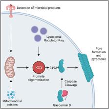
          Graphical Abstract

          In brief

          Devant et al. investigate the actions of reactive oxygen species on the pore-forming protein GSDMD. They show that oxidation of a specific cysteine in GSDMD (C192) promotes its oligomerization, pore-forming activity, and pyroptosis. These findings reveal GSDMD as a redox-regulated protein that connects cellular redox state to inflammatory cell death.

          Related collections

          Most cited references41

          • Record: found
          • Abstract: found
          • Article: not found

          Cleavage of GSDMD by inflammatory caspases determines pyroptotic cell death.

          Inflammatory caspases (caspase-1, -4, -5 and -11) are critical for innate defences. Caspase-1 is activated by ligands of various canonical inflammasomes, and caspase-4, -5 and -11 directly recognize bacterial lipopolysaccharide, both of which trigger pyroptosis. Despite the crucial role in immunity and endotoxic shock, the mechanism for pyroptosis induction by inflammatory caspases is unknown. Here we identify gasdermin D (Gsdmd) by genome-wide clustered regularly interspaced palindromic repeat (CRISPR)-Cas9 nuclease screens of caspase-11- and caspase-1-mediated pyroptosis in mouse bone marrow macrophages. GSDMD-deficient cells resisted the induction of pyroptosis by cytosolic lipopolysaccharide and known canonical inflammasome ligands. Interleukin-1β release was also diminished in Gsdmd(-/-) cells, despite intact processing by caspase-1. Caspase-1 and caspase-4/5/11 specifically cleaved the linker between the amino-terminal gasdermin-N and carboxy-terminal gasdermin-C domains in GSDMD, which was required and sufficient for pyroptosis. The cleavage released the intramolecular inhibition on the gasdermin-N domain that showed intrinsic pyroptosis-inducing activity. Other gasdermin family members were not cleaved by inflammatory caspases but shared the autoinhibition; gain-of-function mutations in Gsdma3 that cause alopecia and skin defects disrupted the autoinhibition, allowing its gasdermin-N domain to trigger pyroptosis. These findings offer insight into inflammasome-mediated immunity/diseases and also change our understanding of pyroptosis and programmed necrosis.
            Bookmark
            • Record: found
            • Abstract: found
            • Article: not found

            Reactive oxygen species (ROS) as pleiotropic physiological signalling agents

            'Reactive oxygen species' (ROS) is an umbrella term for an array of derivatives of molecular oxygen that occur as a normal attribute of aerobic life. Elevated formation of the different ROS leads to molecular damage, denoted as 'oxidative distress'. Here we focus on ROS at physiological levels and their central role in redox signalling via different post-translational modifications, denoted as 'oxidative eustress'. Two species, hydrogen peroxide (H2O2) and the superoxide anion radical (O2·-), are key redox signalling agents generated under the control of growth factors and cytokines by more than 40 enzymes, prominently including NADPH oxidases and the mitochondrial electron transport chain. At the low physiological levels in the nanomolar range, H2O2 is the major agent signalling through specific protein targets, which engage in metabolic regulation and stress responses to support cellular adaptation to a changing environment and stress. In addition, several other reactive species are involved in redox signalling, for instance nitric oxide, hydrogen sulfide and oxidized lipids. Recent methodological advances permit the assessment of molecular interactions of specific ROS molecules with specific targets in redox signalling pathways. Accordingly, major advances have occurred in understanding the role of these oxidants in physiology and disease, including the nervous, cardiovascular and immune systems, skeletal muscle and metabolic regulation as well as ageing and cancer. In the past, unspecific elimination of ROS by use of low molecular mass antioxidant compounds was not successful in counteracting disease initiation and progression in clinical trials. However, controlling specific ROS-mediated signalling pathways by selective targeting offers a perspective for a future of more refined redox medicine. This includes enzymatic defence systems such as those controlled by the stress-response transcription factors NRF2 and nuclear factor-κB, the role of trace elements such as selenium, the use of redox drugs and the modulation of environmental factors collectively known as the exposome (for example, nutrition, lifestyle and irradiation).
              Bookmark
              • Record: found
              • Abstract: found
              • Article: not found

              Mitochondrial reactive oxygen species (ROS) and ROS-induced ROS release.

              Byproducts of normal mitochondrial metabolism and homeostasis include the buildup of potentially damaging levels of reactive oxygen species (ROS), Ca(2+), etc., which must be normalized. Evidence suggests that brief mitochondrial permeability transition pore (mPTP) openings play an important physiological role maintaining healthy mitochondria homeostasis. Adaptive and maladaptive responses to redox stress may involve mitochondrial channels such as mPTP and inner membrane anion channel (IMAC). Their activation causes intra- and intermitochondrial redox-environment changes leading to ROS release. This regenerative cycle of mitochondrial ROS formation and release was named ROS-induced ROS release (RIRR). Brief, reversible mPTP opening-associated ROS release apparently constitutes an adaptive housekeeping function by the timely release from mitochondria of accumulated potentially toxic levels of ROS (and Ca(2+)). At higher ROS levels, longer mPTP openings may release a ROS burst leading to destruction of mitochondria, and if propagated from mitochondrion to mitochondrion, of the cell itself. The destructive function of RIRR may serve a physiological role by removal of unwanted cells or damaged mitochondria, or cause the pathological elimination of vital and essential mitochondria and cells. The adaptive release of sufficient ROS into the vicinity of mitochondria may also activate local pools of redox-sensitive enzymes involved in protective signaling pathways that limit ischemic damage to mitochondria and cells in that area. Maladaptive mPTP- or IMAC-related RIRR may also be playing a role in aging. Because the mechanism of mitochondrial RIRR highlights the central role of mitochondria-formed ROS, we discuss all of the known ROS-producing sites (shown in vitro) and their relevance to the mitochondrial ROS production in vivo. Copyright © 2014 the American Physiological Society.
                Bookmark

                Author and article information

                Journal
                101573691
                39703
                Cell Rep
                Cell Rep
                Cell reports
                2211-1247
                9 February 2023
                31 January 2023
                19 January 2023
                23 February 2023
                : 42
                : 1
                : 112008
                Affiliations
                [1 ]Division of Gastroenterology, Boston Children’s Hospital and Harvard Medical School, 300 Longwood Avenue, Boston, MA 02115, USA
                [2 ]Department of Synthetic Biology and Immunology, National Institute of Chemistry, Hajdrihova 19, 1000 Ljubljana, Slovenia
                [3 ]Graduate School of Biomedicine, University of Ljubljana, Kongresni trg 12, 1000 Ljubljana, Slovenia
                [4 ]EN-FIST Centre of Excellence, Trg Osvobodilne fronte 13, 1000 Ljubljana, Slovenia
                [5 ]Department of Cancer Biology, Dana-Farber Cancer Institute, Boston, MA, USA
                [6 ]Department of Cell Biology, Harvard Medical School, Boston, MA, USA
                [7 ]Ragon Institute of MGH, MIT and Harvard, 400 Technology Square, Cambridge, MA 02139, USA
                [8 ]These authors contributed equally
                [9 ]Lead contact
                Author notes

                AUTHOR CONTRIBUTIONS

                Conceptualization: C.L.E., P.D., I.H.-B., and J.C.K.; methodology, P.D., E.M.N., E.B., C.L.E., I.H.-B., J.R.T., E.T.C., H.X., and J.C.K.; investigation, P.D., E.B., E.M.N., C.L.E., I.H.-B., and H.X.; visualization, P.D., E.B. C.L.E., I.H.-B., E.M.N., and J.R.T.; writing – original draft, P.D. and C.L.E.; writing – review & editing, all authors; funding acquisition, C.L.E., I.H.-B., P.D., A.H.S., J. R.T., E.T.C., H.X., and J.C.K.; supervision, C.L.E., I.H.-B., E.T.C., J.R.T., and J.C.K.

                Article
                NIHMS1870539
                10.1016/j.celrep.2023.112008
                9947919
                36662620
                a61be763-1666-4e09-8b6e-d6a7bd042af5

                This is an open access article under the CC BY-NC-ND license ( http://creativecommons.org/licenses/by-nc-nd/4.0/).

                History
                Categories
                Article

                Cell biology
                Cell biology

                Comments

                Comment on this article