20
views
0
recommends
+1 Recommend
0 collections
    0
    shares
      • Record: found
      • Abstract: found
      • Article: found
      Is Open Access

      Tumor resistance to ferroptosis driven by Stearoyl-CoA Desaturase-1 (SCD1) in cancer cells and Fatty Acid Biding Protein-4 (FABP4) in tumor microenvironment promote tumor recurrence

      research-article

      Read this article at

      Bookmark
          There is no author summary for this article yet. Authors can add summaries to their articles on ScienceOpen to make them more accessible to a non-specialist audience.

          Abstract

          Problem

          Tumor recurrence is a major clinical issue that represents the principal cause of cancer-related deaths, with few targetable common pathways. Mechanisms by which residual tumors persist and progress under a continuous shift between hypoxia-reoxygenation after neoadjuvent-therapy are unknown. In this study, we investigated the role of lipid metabolism and tumor redox balance in tumor recurrence.

          Methods

          Lipidomics, proteomics and mass spectrometry imaging approaches where applied to mouse tumor models of recurrence. Genetic and pharmacological inhibitions of lipid mediators in tumors were used in vivo and in functional assays in vitro.

          Results

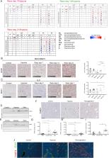
          We found that stearoyl-CoA desaturase-1 (SCD1) expressed by cancer cells and fatty acid binding protein-4 (FABP4) produced by tumor endothelial cells (TECs) and adipocytes in the tumor microenvironment (TME) are essential for tumor relapse in response to tyrosine kinase inhibitors (TKI) and chemotherapy. SCD1 and FABP4 were also found upregulated in recurrent human breast cancer samples and correlated with worse prognosis of cancer patients with different types of tumors. Mechanistically, SCD1 leads to fatty acid (FA) desaturation and FABP4 derived from TEM enhances lipid droplet (LD) in cancer cells, which cooperatively protect from oxidative stress-induced ferroptosis. We revealed that lipid mobilization and desaturation elicit tumor intrinsic antioxidant and anti-ferroptotic resources for survival and regrowth in a harsh TME. Inhibition of lipid transport from TME by FABP4 inhibitor reduced tumor regrowth and by genetic — or by pharmacological — targeting SCD1 in vivo, tumor regrowth was abolished completely.

          Conclusion

          This finding unveils that it is worth taking advantage of tumor lipid addiction, as a tumor vulnerability to design novel treatment strategy to prevent cancer recurrence.

          Graphical abstract

          Highlights

          • Increased oxidative stress markers and lipid metabolism in residual tumors.

          • Expression of SCD1 in cancer cells and FABP4 in the tumor microenvironment drive tumor recurrence.

          • Fatty acid desaturation by SCD1 and lipid transport by FABP4 confer resistance to ROS and ferroptosis.

          • Blocking SCD1 and FABP4 sensitized cancer cells to ROS-induced ferroptosis and reduced tumor recurrence.

          Related collections

          Most cited references48

          • Record: found
          • Abstract: found
          • Article: not found

          Ferroptosis: an iron-dependent form of nonapoptotic cell death.

          Nonapoptotic forms of cell death may facilitate the selective elimination of some tumor cells or be activated in specific pathological states. The oncogenic RAS-selective lethal small molecule erastin triggers a unique iron-dependent form of nonapoptotic cell death that we term ferroptosis. Ferroptosis is dependent upon intracellular iron, but not other metals, and is morphologically, biochemically, and genetically distinct from apoptosis, necrosis, and autophagy. We identify the small molecule ferrostatin-1 as a potent inhibitor of ferroptosis in cancer cells and glutamate-induced cell death in organotypic rat brain slices, suggesting similarities between these two processes. Indeed, erastin, like glutamate, inhibits cystine uptake by the cystine/glutamate antiporter (system x(c)(-)), creating a void in the antioxidant defenses of the cell and ultimately leading to iron-dependent, oxidative death. Thus, activation of ferroptosis results in the nonapoptotic destruction of certain cancer cells, whereas inhibition of this process may protect organisms from neurodegeneration. Copyright © 2012 Elsevier Inc. All rights reserved.
            Bookmark
            • Record: found
            • Abstract: found
            • Article: not found

            Regulation of ferroptotic cancer cell death by GPX4.

            Ferroptosis is a form of nonapoptotic cell death for which key regulators remain unknown. We sought a common mediator for the lethality of 12 ferroptosis-inducing small molecules. We used targeted metabolomic profiling to discover that depletion of glutathione causes inactivation of glutathione peroxidases (GPXs) in response to one class of compounds and a chemoproteomics strategy to discover that GPX4 is directly inhibited by a second class of compounds. GPX4 overexpression and knockdown modulated the lethality of 12 ferroptosis inducers, but not of 11 compounds with other lethal mechanisms. In addition, two representative ferroptosis inducers prevented tumor growth in xenograft mouse tumor models. Sensitivity profiling in 177 cancer cell lines revealed that diffuse large B cell lymphomas and renal cell carcinomas are particularly susceptible to GPX4-regulated ferroptosis. Thus, GPX4 is an essential regulator of ferroptotic cancer cell death. Copyright © 2014 Elsevier Inc. All rights reserved.
              Bookmark
              • Record: found
              • Abstract: found
              • Article: not found

              Peroxidation of polyunsaturated fatty acids by lipoxygenases drives ferroptosis.

              Ferroptosis is form of regulated nonapoptotic cell death that is involved in diverse disease contexts. Small molecules that inhibit glutathione peroxidase 4 (GPX4), a phospholipid peroxidase, cause lethal accumulation of lipid peroxides and induce ferroptotic cell death. Although ferroptosis has been suggested to involve accumulation of reactive oxygen species (ROS) in lipid environments, the mediators and substrates of ROS generation and the pharmacological mechanism of GPX4 inhibition that generates ROS in lipid environments are unknown. We report here the mechanism of lipid peroxidation during ferroptosis, which involves phosphorylase kinase G2 (PHKG2) regulation of iron availability to lipoxygenase enzymes, which in turn drive ferroptosis through peroxidation of polyunsaturated fatty acids (PUFAs) at the bis-allylic position; indeed, pretreating cells with PUFAs containing the heavy hydrogen isotope deuterium at the site of peroxidation (D-PUFA) prevented PUFA oxidation and blocked ferroptosis. We further found that ferroptosis inducers inhibit GPX4 by covalently targeting the active site selenocysteine, leading to accumulation of PUFA hydroperoxides. In summary, we found that PUFA oxidation by lipoxygenases via a PHKG2-dependent iron pool is necessary for ferroptosis and that the covalent inhibition of the catalytic selenocysteine in Gpx4 prevents elimination of PUFA hydroperoxides; these findings suggest new strategies for controlling ferroptosis in diverse contexts.
                Bookmark

                Author and article information

                Contributors
                Journal
                Redox Biol
                Redox Biol
                Redox Biology
                Elsevier
                2213-2317
                14 May 2021
                July 2021
                14 May 2021
                : 43
                : 102006
                Affiliations
                [a ]Laboratory of Biology of Tumor and Developmental Biology, GIGA Cancer, Liège University, Liège, Belgium
                [b ]Cancer Metabolism and Tumor Microenvironment Group, GIGA Cancer, Liège University, Liège, Belgium
                [c ]Division of Gastroenterology, Department of Internal Medicine, Kobe University Graduate School of Medicine, Kobe, Japan
                [d ]Mass Spectrometry Laboratory, MolSys Research Unit, University of Liège, Liège, Belgium
                [e ]Department of Clinical Pharmacology and Pharmacoepidemiology, Heidelberg University Hospital, Heidelberg, Germany
                [f ]Metastasis Research Laboratory, GIGA Cancer, University of Liège, 4000, Liège, Belgium
                [g ]Department of Omics Medicine, Hyogo College of Medicine, Nishinomiya, Japan
                [h ]Division of Metabolomics Research, Department of Internal Related, Kobe University Graduate School of Medicine, Kobe, Japan
                [i ]AMED-CREST, AMED, Kobe, Japan
                [j ]Clinical Research Unit, Fondation Hôpitaux Robert Schuman, Hôpitaux Robert Schuman, Luxembourg
                Author notes
                []Corresponding author. Cancer Metabolism and Tumor Microenvironment Group, Laboratory of Tumor and Developmental Biology, GIGA-cancer, Liège University, Liège, Belgium. nesounni@ 123456uliege.be
                [1]

                These authors contributed equally.

                Article
                S2213-2317(21)00164-6 102006
                10.1016/j.redox.2021.102006
                8163990
                34030117
                a569996f-1206-49e3-8a69-60dda7252288
                © 2021 The Authors

                This is an open access article under the CC BY-NC-ND license (http://creativecommons.org/licenses/by-nc-nd/4.0/).

                History
                : 9 April 2021
                : 4 May 2021
                : 10 May 2021
                Categories
                Research Paper

                lipid metabolism,hypoxia,reoxygenation,drug-resistance,tumor-microenvironment,ros-ferroptosis

                Comments

                Comment on this article