31
views
0
recommends
+1 Recommend
0 collections
    0
    shares
      • Record: found
      • Abstract: found
      • Article: found
      Is Open Access

      Tannic Acid Modified Silver Nanoparticles Show Antiviral Activity in Herpes Simplex Virus Type 2 Infection

      research-article

      Read this article at

      Bookmark
          There is no author summary for this article yet. Authors can add summaries to their articles on ScienceOpen to make them more accessible to a non-specialist audience.

          Abstract

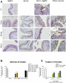
          The interaction between silver nanoparticles and herpesviruses is attracting great interest due to their antiviral activity and possibility to use as microbicides for oral and anogenital herpes. In this work, we demonstrate that tannic acid modified silver nanoparticles sized 13 nm, 33 nm and 46 nm are capable of reducing HSV-2 infectivity both in vitro and in vivo. The antiviral activity of tannic acid modified silver nanoparticles was size-related, required direct interaction and blocked virus attachment, penetration and further spread. All tested tannic acid modified silver nanoparticles reduced both infection and inflammatory reaction in the mouse model of HSV-2 infection when used at infection or for a post-infection treatment. Smaller-sized nanoparticles induced production of cytokines and chemokines important for anti-viral response. The corresponding control buffers with tannic acid showed inferior antiviral effects in vitro and were ineffective in blocking in vivo infection. Our results show that tannic acid modified silver nanoparticles are good candidates for microbicides used in treatment of herpesvirus infections.

          Related collections

          Most cited references50

          • Record: found
          • Abstract: found
          • Article: not found

          Does the antibacterial activity of silver nanoparticles depend on the shape of the nanoparticle? A study of the Gram-negative bacterium Escherichia coli.

          In this work we investigated the antibacterial properties of differently shaped silver nanoparticles against the gram-negative bacterium Escherichia coli, both in liquid systems and on agar plates. Energy-filtering transmission electron microscopy images revealed considerable changes in the cell membranes upon treatment, resulting in cell death. Truncated triangular silver nanoplates with a {111} lattice plane as the basal plane displayed the strongest biocidal action, compared with spherical and rod-shaped nanoparticles and with Ag(+) (in the form of AgNO(3)). It is proposed that nanoscale size and the presence of a {111} plane combine to promote this biocidal property. To our knowledge, this is the first comparative study on the bactericidal properties of silver nanoparticles of different shapes, and our results demonstrate that silver nanoparticles undergo a shape-dependent interaction with the gram-negative organism E. coli.
            Bookmark
            • Record: found
            • Abstract: found
            • Article: found
            Is Open Access

            Interaction of silver nanoparticles with HIV-1

            The interaction of nanoparticles with biomolecules and microorganisms is an expanding field of research. Within this field, an area that has been largely unexplored is the interaction of metal nanoparticles with viruses. In this work, we demonstrate that silver nanoparticles undergo a size-dependent interaction with HIV-1, with nanoparticles exclusively in the range of 1–10 nm attached to the virus. The regular spatial arrangement of the attached nanoparticles, the center-to-center distance between nanoparticles, and the fact that the exposed sulfur-bearing residues of the glycoprotein knobs would be attractive sites for nanoparticle interaction suggest that silver nanoparticles interact with the HIV-1 virus via preferential binding to the gp120 glycoprotein knobs. Due to this interaction, silver nanoparticles inhibit the virus from binding to host cells, as demonstrated in vitro.
              Bookmark
              • Record: found
              • Abstract: not found
              • Article: not found

              A review of the in vivo and in vitro toxicity of silver and gold particulates: particle attributes and biological mechanisms responsible for the observed toxicity.

              This review is concerned with evaluating the toxicity associated with human exposure to silver and gold nanoparticles (NPs), due to the relative abundance of toxicity data available for these particles, when compared to other metal particulates. This has allowed knowledge on the current understanding of the field to be gained, and has demonstrated where gaps in knowledge are. It is anticipated that evaluating the hazards associated with silver and gold particles will ultimately enable risk assessments to be completed, by combining this information with knowledge on the level of human exposure. The quantity of available hazard information for metals is greatest for silver particulates, due to its widespread inclusion within a number of diverse products (including clothes and wound dressings), which primarily arises from its antibacterial behaviour. Gold has been used on numerous occasions to assess the biodistribution and cellular uptake of NPs following exposure. Inflammatory, oxidative, genotoxic, and cytotoxic consequences are associated with silver particulate exposure, and are inherently linked. The primary site of gold and silver particulate accumulation has been consistently demonstrated to be the liver, and it is therefore relevant that a number of in vitro investigations have focused on this potential target organ. However, in general there is a lack of in vivo and in vitro toxicity information that allows correlations between the findings to be made. Instead a focus on the tissue distribution of particles following exposure is evident within the available literature, which can be useful in directing appropriate in vitro experimentation by revealing potential target sites of toxicity. The experimental design has the potential to impact on the toxicological observations, and in particular the use of excessively high particle concentrations has been observed. As witnessed for other particle types, gold and silver particle sizes are influential in dictating the observed toxicity, with smaller particles exhibiting a greater response than their larger counterparts, and this is likely to be driven by differences in particle surface area, when administered at an equal-mass dose. A major obstacle, at present, is deciphering whether the responses related to silver nanoparticulate exposure derive from their small size, or particle dissolution contributes to the observed toxicity. Alternatively, a combination of both may be responsible, as the release of ions would be expected to be greater for smaller particles.
                Bookmark

                Author and article information

                Contributors
                Role: Editor
                Journal
                PLoS One
                PLoS ONE
                plos
                plosone
                PLoS ONE
                Public Library of Science (San Francisco, USA )
                1932-6203
                2014
                12 August 2014
                : 9
                : 8
                : e104113
                Affiliations
                [1 ]Department of Regenerative Medicine, Military Institute of Hygiene and Epidemiology, Warsaw, Poland
                [2 ]Department of Materials Technology and Chemistry, Faculty of Chemistry, University of Lodz, Lodz, Poland
                [3 ]Faculty of Chemistry, University of Warsaw, Warsaw, Poland
                [4 ]Department of Preclinical Sciences, Faculty of Veterinary Medicine, Warsaw University of Life Sciences, Warsaw, Poland
                [5 ]Laboratory of Electron and Confocal Microscopy, Faculty of Biology, University of Warsaw, Warsaw, Poland
                [6 ]Department of Morphological Sciences, Faculty of Veterinary Medicine, Warsaw University of Life Sciences, Warsaw, Poland
                [7 ]Department of Genetics and Animal Breeding, Faculty of Animal Science, Warsaw University of Life Sciences, Warsaw, Poland
                Cincinnati Childrens Hospital Medical Center, United States of America
                Author notes

                Competing Interests: The authors have declared that no competing interests exist.

                Conceived and designed the experiments: MK GC JG. Performed the experiments: MK PB PO ET MG GC. Analyzed the data: PO MG JG MK. Contributed reagents/materials/analysis tools: JN JS MD GC JG ZN. Contributed to the writing of the manuscript: MK PO JG GC. Obtained agreement for the transfer of 291.03C cell line for academic or research institution from Oregon Health and Science University: MK.

                Article
                PONE-D-14-16402
                10.1371/journal.pone.0104113
                4130517
                25117537
                a4acc2c1-b4b0-4f6a-8e3c-a5568d4dbbaf
                Copyright @ 2014

                This is an open-access article distributed under the terms of the Creative Commons Attribution License, which permits unrestricted use, distribution, and reproduction in any medium, provided the original author and source are credited.

                History
                : 11 April 2014
                : 7 July 2014
                Page count
                Pages: 15
                Funding
                This work was supported by the Polish National Science Centre grant No. 2011/03/B/NZ6/04878 (for MK) and Centre for Preclinical Research and Technology (CePT) Project No. POIG.02.02.00-14-024/08-0 (for MG and MD). The funders had no role in study design, data collection and analysis, decision to publish, or preparation of the manuscript.
                Categories
                Research Article
                Biology and Life Sciences
                Microbiology
                Virology
                Engineering and Technology
                Nanotechnology
                Nanoparticles
                Medicine and Health Sciences
                Women's Health
                Obstetrics and Gynecology
                Gynecologic Infections
                Custom metadata
                The authors confirm that all data underlying the findings are fully available without restriction. All relevant data are within the paper and its Supporting Information files.

                Uncategorized
                Uncategorized

                Comments

                Comment on this article