2
views
0
recommends
+1 Recommend
0 collections
    0
    shares
      • Record: found
      • Abstract: found
      • Article: found
      Is Open Access

      Intracellular calcium links milk stasis to lysosome-dependent cell death during early mammary gland involution

      research-article

      Read this article at

      Bookmark
          There is no author summary for this article yet. Authors can add summaries to their articles on ScienceOpen to make them more accessible to a non-specialist audience.

          Abstract

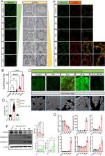
          Involution of the mammary gland after lactation is a dramatic example of coordinated cell death. Weaning causes distension of the alveolar structures due to the accumulation of milk, which, in turn, activates STAT3 and initiates a caspase-independent but lysosome-dependent cell death (LDCD) pathway. Although the importance of STAT3 and LDCD in early mammary involution is well established, it has not been entirely clear how milk stasis activates STAT3. In this report, we demonstrate that protein levels of the PMCA2 calcium pump are significantly downregulated within 2–4 h of experimental milk stasis. Reductions in PMCA2 expression correlate with an increase in cytoplasmic calcium in vivo as measured by multiphoton intravital imaging of GCaMP6f fluorescence. These events occur concomitant with the appearance of nuclear pSTAT3 expression but prior to significant activation of LDCD or its previously implicated mediators such as LIF, IL6, and TGFβ3, all of which appear to be upregulated by increased intracellular calcium. We further demonstrate that increased intracellular calcium activates STAT3 by inducing degradation of its negative regulator, SOCS3. We also observed that milk stasis, loss of PMCA2 expression and increased intracellular calcium levels activate TFEB, an important regulator of lysosome biogenesis through a process involving inhibition of CDK4/6 and cell cycle progression. In summary, these data suggest that intracellular calcium serves as an important proximal biochemical signal linking milk stasis to STAT3 activation, increased lysosomal biogenesis, and lysosome-mediated cell death.

          Supplementary Information

          The online version contains supplementary material available at 10.1007/s00018-023-05044-8.

          Related collections

          Most cited references63

          • Record: found
          • Abstract: found
          • Article: found
          Is Open Access

          Moderated estimation of fold change and dispersion for RNA-seq data with DESeq2

          In comparative high-throughput sequencing assays, a fundamental task is the analysis of count data, such as read counts per gene in RNA-seq, for evidence of systematic changes across experimental conditions. Small replicate numbers, discreteness, large dynamic range and the presence of outliers require a suitable statistical approach. We present DESeq2, a method for differential analysis of count data, using shrinkage estimation for dispersions and fold changes to improve stability and interpretability of estimates. This enables a more quantitative analysis focused on the strength rather than the mere presence of differential expression. The DESeq2 package is available at http://www.bioconductor.org/packages/release/bioc/html/DESeq2.html. Electronic supplementary material The online version of this article (doi:10.1186/s13059-014-0550-8) contains supplementary material, which is available to authorized users.
            Bookmark
            • Record: found
            • Abstract: found
            • Article: not found

            HISAT: a fast spliced aligner with low memory requirements.

            HISAT (hierarchical indexing for spliced alignment of transcripts) is a highly efficient system for aligning reads from RNA sequencing experiments. HISAT uses an indexing scheme based on the Burrows-Wheeler transform and the Ferragina-Manzini (FM) index, employing two types of indexes for alignment: a whole-genome FM index to anchor each alignment and numerous local FM indexes for very rapid extensions of these alignments. HISAT's hierarchical index for the human genome contains 48,000 local FM indexes, each representing a genomic region of ∼64,000 bp. Tests on real and simulated data sets showed that HISAT is the fastest system currently available, with equal or better accuracy than any other method. Despite its large number of indexes, HISAT requires only 4.3 gigabytes of memory. HISAT supports genomes of any size, including those larger than 4 billion bases.
              Bookmark
              • Record: found
              • Abstract: found
              • Article: found
              Is Open Access

              clusterProfiler 4.0: A universal enrichment tool for interpreting omics data

              Summary Functional enrichment analysis is pivotal for interpreting high-throughput omics data in life science. It is crucial for this type of tool to use the latest annotation databases for as many organisms as possible. To meet these requirements, we present here an updated version of our popular Bioconductor package, clusterProfiler 4.0. This package has been enhanced considerably compared with its original version published 9 years ago. The new version provides a universal interface for functional enrichment analysis in thousands of organisms based on internally supported ontologies and pathways as well as annotation data provided by users or derived from online databases. It also extends the dplyr and ggplot2 packages to offer tidy interfaces for data operation and visualization. Other new features include gene set enrichment analysis and comparison of enrichment results from multiple gene lists. We anticipate that clusterProfiler 4.0 will be applied to a wide range of scenarios across diverse organisms.
                Bookmark

                Author and article information

                Contributors
                jaekwang.jeong@yale.edu
                john.wysolmerski@yale.edu
                Journal
                Cell Mol Life Sci
                Cell Mol Life Sci
                Cellular and Molecular Life Sciences
                Springer International Publishing (Cham )
                1420-682X
                1420-9071
                12 January 2024
                12 January 2024
                2024
                : 81
                : 1
                : 29
                Affiliations
                [1 ]Section of Endocrinology and Metabolism, Department of Internal Medicine, Yale University School of Medicine, ( https://ror.org/03v76x132) New Haven, CT USA
                [2 ]GRID grid.222754.4, ISNI 0000 0001 0840 2678, Department of Biomedical Sciences, , Korea University College of Medicine, ; Seoul, Republic of Korea
                [3 ]Departments of Cell Biology and of Neuroscience, Wu Tsai Institute, Yale University School of Medicine, ( https://ror.org/03v76x132) New Haven, CT 06510 USA
                [4 ]Division of Phamacology, School of Korean Medicine, Pusan National University, ( https://ror.org/01an57a31) Yangsan, Gyeongnam 50612 Republic of Korea
                [5 ]GRID grid.47100.32, ISNI 0000000419368710, Department of Genetics, , Yale School of Medicine, ; New Haven, CT 06510 USA
                [6 ]Integrated Science Engineering Division, Underwood International College, Yonsei University, ( https://ror.org/01wjejq96) Seoul, Republic of Korea
                [7 ]GRID grid.47100.32, ISNI 0000000419368710, Departments of Immunobiology and Laboratory Medicine, , Yale School of Medicine, ; New Haven, CT 06510 USA
                [8 ]GRID grid.15444.30, ISNI 0000 0004 0470 5454, Department of Biomedical Sciences, Graduate School of Medical Science, Brain Korea 21 Project, Gangnam Severance Hospital, , Yonsei University College of Medicine, ; Seoul, 06230 Republic of Korea
                Author information
                http://orcid.org/0000-0001-8865-2545
                Article
                5044
                10.1007/s00018-023-05044-8
                10784359
                38212474
                a46783ea-c236-42a8-9614-66b3870d8cc4
                © The Author(s) 2024

                Open Access This article is licensed under a Creative Commons Attribution 4.0 International License, which permits use, sharing, adaptation, distribution and reproduction in any medium or format, as long as you give appropriate credit to the original author(s) and the source, provide a link to the Creative Commons licence, and indicate if changes were made. The images or other third party material in this article are included in the article's Creative Commons licence, unless indicated otherwise in a credit line to the material. If material is not included in the article's Creative Commons licence and your intended use is not permitted by statutory regulation or exceeds the permitted use, you will need to obtain permission directly from the copyright holder. To view a copy of this licence, visit http://creativecommons.org/licenses/by/4.0/.

                History
                : 10 June 2023
                : 17 October 2023
                : 7 November 2023
                Funding
                Funded by: FundRef http://dx.doi.org/10.13039/100000002, National Institutes of Health;
                Award ID: RO1 GM105718
                Award ID: R01 HD100468
                Award ID: R01 HD076248
                Award Recipient :
                Funded by: NATIONAL RESEARCH FOUNDATION OF KOREA
                Award ID: 2018R1C1B6002803
                Award ID: 2022R1A4A2000827
                Award Recipient :
                Categories
                Original Article
                Custom metadata
                © Springer Nature Switzerland AG 2024

                Molecular biology
                ldcd,calcium,pmca2,stat3,tfeb
                Molecular biology
                ldcd, calcium, pmca2, stat3, tfeb

                Comments

                Comment on this article