3
views
0
recommends
+1 Recommend
0 collections
    0
    shares
      • Record: found
      • Abstract: found
      • Article: found
      Is Open Access

      Juvenile trabecular ossifying fibroma: A case of extensive lesion of the maxilla

      case-report

      Read this article at

      Bookmark
          There is no author summary for this article yet. Authors can add summaries to their articles on ScienceOpen to make them more accessible to a non-specialist audience.

          Abstract

          Introduction and importance

          Juvenile trabecular ossifying fibroma is a rare benign tumor of childhood affecting the facial bones rarely described in literature. Its aggressive growth and high tendency of recurrence make it a real challenge for diagnosis and care.

          Case presentation

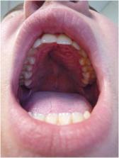
          This article presents a case of an extensive juvenile trabecular ossifying fibroma of the maxilla in a 14-year-old boy, which required surgical intervention with immediate bone reconstruction using an autogenous graft (iliac crest).

          No recurrence has been found after two and a half years of follow-up.

          Clinical discussion

          Common symptoms of juvenile trabecular ossifying fibroma include painless swelling, facial asymmetry, jaw deformity and teeth displacement. Differential diagnosis should consider other forms like psammomatoid ossifying fibroma, osteofibrous dysplasia, fibrous dysplasia, ameloblastoma, odontoma, or even poorly differentiated carcinoma.

          Conclusion

          Currently, there is no recommendation or consensus for the treatment of juvenile trabecular ossifying fibroma. The objective of treatment is also to preserve growth and development and conserve the nervous structure and the function, as mastication, vision.

          Early diagnosis and appropriate care are essential to prevent morphological and functional defects in young patients. Regular and long-term follow-up is essential due to the high recurrence rate.

          Highlight

          • This case report present a rare benign tumor of childhood affecting the facial bones with aggressive growth with aesthetic and functional sequelae.

          • This pathology, which is rarely found in the literature, requires rapid treatment and regular follow-up because recurrences are frequent.

          • The purpose of this article is to expose a case of extensive trabecular juvenile ossifying fibroma of the maxilla in a 14 years old boy that required surgical intervention with bone reconstruction and which we were able to survey for two and a half years.

          Related collections

          Most cited references21

          • Record: found
          • Abstract: found
          • Article: not found

          The SCARE 2020 Guideline: Updating Consensus Surgical CAse REport (SCARE) Guidelines

          The SCARE Guidelines were first published in 2016 and were last updated in 2018. They provide a structure for reporting surgical case reports and are used and endorsed by authors, journal editors and reviewers, in order to increase robustness and transparency in reporting surgical cases. They must be kept up to date in order to drive forwards reporting quality. As such, we have updated these guidelines via a DELPHI consensus exercise.
            Bookmark
            • Record: found
            • Abstract: found
            • Article: not found

            Psammomatoid and trabecular juvenile ossifying fibroma of the craniofacial skeleton: two distinct clinicopathologic entities.

            The term juvenile ossifying fibroma is used in the literature in naming 2 microscopically distinct fibro-osseous lesions of the craniofacial skeleton. One is characterized by small uniform spherical ossicles resembling psammoma bodies (psammomatoid juvenile ossifying fibroma). The other is distinguished by trabeculae of fibrillary osteoid and woven bone (trabecular juvenile ossifying fibroma). Three new cases of each type are reported, and the literature is extensively reviewed for published cases of these 2 entities.Psammomatoid juvenile ossifying fibroma is reported more commonly than trabecular juvenile ossifying fibroma. It affects patients from a wider age range (3 months to 72 years) and an older mean age range (16-33 years) as compared with 2 to 12 years and 8(1/2) to 12 years, respectively, for trabecular juvenile ossifying fibroma. In both types there is a slight male predominance and the lesions are unencapsulated and tend to infiltrate adjacent bone. A significant difference between the 2 tumors is their site of occurrence. Although psammomatoid juvenile ossifying fibroma occurs predominantly in the sinonasal and orbital bones, trabecular juvenile ossifying fibroma predominantly affects the jaws. Aggressive growth occurs in some-but not all-cases of both types. Such behavior may be related to younger patient age and the concurrent development of aneurysmal bone cysts, which is seen more frequently in psammomatoid juvenile ossifying fibroma. This study demonstrates that not only histologic but also demographic and clinical differences between psammomatoid juvenile ossifying fibroma and trabecular juvenile ossifying fibroma warrant their classification as 2 distinct clinicopathologic entities.
              Bookmark
              • Record: found
              • Abstract: found
              • Article: not found

              Fibro-osseous lesions of the craniofacial skeleton: an update.

              Benign fibro-osseous lesions of the craniofacial skeleton (BFOL) are a variant group of intraosseous disease processes that share similar microscopic features characterized by hypercellular fibroblastic stroma containing various combinations of bone or cementum-like tissue and other calcified structures [1-6]. Whereas some are diagnosable histologically, most require a combined assessment of clinical, microscopic and radiologic features. Some BFOL of the craniofacial complex are unique to that location whereas others are encountered in bones from other regions. Reactive, neoplastic, developmental and dysplastic pathologic processes are included under the rubric of BFOL and treatment varies from disease to disease. This review will discuss the clinical, microscopic and radiologic aspects of the more important types of BFOL of the craniofacial complex with updated information on underlying genetic and molecular pathogenic mechanisms of disease. Four main groups of BFOLs will be addressed.
                Bookmark

                Author and article information

                Contributors
                Journal
                Int J Surg Case Rep
                Int J Surg Case Rep
                International Journal of Surgery Case Reports
                Elsevier
                2210-2612
                22 August 2023
                October 2023
                22 August 2023
                : 111
                : 108620
                Affiliations
                [a ]Faculty of Dental Surgery, Nancy, France
                [b ]Dept of anatomopathology, University Hospital, Nancy, France
                [c ]Medical Professor Dept. of Oral and Maxillofacial Surgery, and plastic surgery, University Hospital, Nancy, France
                [d ]Medical Professor Dept. of Oral and Maxillofacial Surgery and plastic surgery, university hospital, Nancy, France
                Author notes
                [* ]Corresponding author at: faculty of dentistry of Lorraine, 7 Av. De la Foret de Haye, 54500 Vandœuvre-Lès-Nancy, France. maeva.nedelec9@ 123456etu.univ-lorraine.fr
                Article
                S2210-2612(23)00749-6 108620
                10.1016/j.ijscr.2023.108620
                10502336
                37703694
                a35201da-3106-4769-a0cc-493a6bbf1be3
                © 2023 Published by Elsevier Ltd on behalf of IJS Publishing Group Ltd.

                This is an open access article under the CC BY-NC-ND license (http://creativecommons.org/licenses/by-nc-nd/4.0/).

                History
                : 23 June 2023
                : 30 July 2023
                : 31 July 2023
                Categories
                Case Report

                case report,trabecular juvenile ossifying fibroma,benign fibro-osseous lesions,maxillofacial bone pathology

                Comments

                Comment on this article