32
views
0
recommends
+1 Recommend
0 collections
    32
    shares
      • Record: found
      • Abstract: found
      • Article: found
      Is Open Access

      A PI3K p110β–Rac signalling loop mediates Pten-loss-induced perturbation of haematopoiesis and leukaemogenesis

      research-article

      Read this article at

      Bookmark
          There is no author summary for this article yet. Authors can add summaries to their articles on ScienceOpen to make them more accessible to a non-specialist audience.

          Abstract

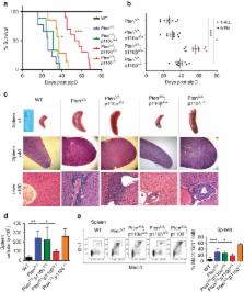
          The tumour suppressor PTEN, which antagonizes PI3K signalling, is frequently inactivated in haematologic malignancies. In mice, deletion of PTEN in haematopoietic stem cells (HSCs) causes perturbed haematopoiesis, myeloproliferative neoplasia (MPN) and leukaemia. Although the roles of the PI3K isoforms have been studied in PTEN-deficient tumours, their individual roles in PTEN-deficient HSCs are unknown. Here we show that when we delete PTEN in HSCs using the Mx1–Cre system, p110β ablation prevents MPN, improves HSC function and suppresses leukaemia initiation. Pharmacologic inhibition of p110β in PTEN-deficient mice recapitulates these genetic findings, but suggests involvement of both Akt-dependent and -independent pathways. Further investigation reveals that a p110β–Rac signalling loop plays a critical role in PTEN-deficient HSCs. Together, these data suggest that myeloid neoplasia driven by PTEN loss is dependent on p110β via p110β–Rac-positive-feedback loop, and that disruption of this loop may offer a new and effective therapeutic strategy for PTEN-deficient leukaemia.

          Abstract

          The tumor suppressor PTEN antagonizes the PI3K signalling pathway and is frequently inactivated in haematological malignancies. Here, the authors unravel the main contribution of the PI3K isoform p110ß to leukemic transformation driven by PTEN-loss.

          Related collections

          Most cited references43

          • Record: found
          • Abstract: found
          • Article: not found

          Prognostic relevance of integrated genetic profiling in acute myeloid leukemia.

          Acute myeloid leukemia (AML) is a heterogeneous disease with respect to presentation and clinical outcome. The prognostic value of recently identified somatic mutations has not been systematically evaluated in a phase 3 trial of treatment for AML. We performed a mutational analysis of 18 genes in 398 patients younger than 60 years of age who had AML and who were randomly assigned to receive induction therapy with high-dose or standard-dose daunorubicin. We validated our prognostic findings in an independent set of 104 patients. We identified at least one somatic alteration in 97.3% of the patients. We found that internal tandem duplication in FLT3 (FLT3-ITD), partial tandem duplication in MLL (MLL-PTD), and mutations in ASXL1 and PHF6 were associated with reduced overall survival (P=0.001 for FLT3-ITD, P=0.009 for MLL-PTD, P=0.05 for ASXL1, and P=0.006 for PHF6); CEBPA and IDH2 mutations were associated with improved overall survival (P=0.05 for CEBPA and P=0.01 for IDH2). The favorable effect of NPM1 mutations was restricted to patients with co-occurring NPM1 and IDH1 or IDH2 mutations. We identified genetic predictors of outcome that improved risk stratification among patients with AML, independently of age, white-cell count, induction dose, and post-remission therapy, and validated the significance of these predictors in an independent cohort. High-dose daunorubicin, as compared with standard-dose daunorubicin, improved the rate of survival among patients with DNMT3A or NPM1 mutations or MLL translocations (P=0.001) but not among patients with wild-type DNMT3A, NPM1, and MLL (P=0.67). We found that DNMT3A and NPM1 mutations and MLL translocations predicted an improved outcome with high-dose induction chemotherapy in patients with AML. These findings suggest that mutational profiling could potentially be used for risk stratification and to inform prognostic and therapeutic decisions regarding patients with AML. (Funded by the National Cancer Institute and others.).
            Bookmark
            • Record: found
            • Abstract: found
            • Article: not found

            Pten dependence distinguishes haematopoietic stem cells from leukaemia-initiating cells.

            Recent advances have highlighted extensive phenotypic and functional similarities between normal stem cells and cancer stem cells. This raises the question of whether disease therapies can be developed that eliminate cancer stem cells without eliminating normal stem cells. Here we address this issue by conditionally deleting the Pten tumour suppressor gene in adult haematopoietic cells. This led to myeloproliferative disease within days and transplantable leukaemias within weeks. Pten deletion also promoted haematopoietic stem cell (HSC) proliferation. However, this led to HSC depletion via a cell-autonomous mechanism, preventing these cells from stably reconstituting irradiated mice. In contrast to leukaemia-initiating cells, HSCs were therefore unable to maintain themselves without Pten. These effects were mostly mediated by mTOR as they were inhibited by rapamycin. Rapamycin not only depleted leukaemia-initiating cells but also restored normal HSC function. Mechanistic differences between normal stem cells and cancer stem cells can thus be targeted to deplete cancer stem cells without damaging normal stem cells.
              Bookmark
              • Record: found
              • Abstract: found
              • Article: not found

              Rational design and characterization of a Rac GTPase-specific small molecule inhibitor.

              The signaling pathways mediated by Rho family GTPases have been implicated in many aspects of cell biology. The specificity of the pathways is achieved in part by the selective interaction between Dbl family guanine nucleotide exchange factors (GEFs) and their Rho GTPase substrates. Here, we report a first-generation small-molecule inhibitor of Rac GTPase targeting Rac activation by GEF. The chemical compound NSC23766 was identified by a structure-based virtual screening of compounds that fit into a surface groove of Rac1 known to be critical for GEF specification. In vitro it could effectively inhibit Rac1 binding and activation by the Rac-specific GEF Trio or Tiam1 in a dose-dependent manner without interfering with the closely related Cdc42 or RhoA binding or activation by their respective GEFs or with Rac1 interaction with BcrGAP or effector PAK1. In cells, it potently blocked serum or platelet-derived growth factor-induced Rac1 activation and lamellipodia formation without affecting the activity of endogenous Cdc42 or RhoA. Moreover, this compound reduced Trio or Tiam1 but not Vav, Lbc, Intersectin, or a constitutively active Rac1 mutant-stimulated cell growth and suppressed Trio, Tiam1, or Ras-induced cell transformation. When applied to human prostate cancer PC-3 cells, it was able to inhibit the proliferation, anchorage-independent growth and invasion phenotypes that require the endogenous Rac1 activity. Thus, NSC23766 constitutes a Rac-specific small-molecule inhibitor that could be useful to study the role of Rac in various cellular functions and to reverse tumor cell phenotypes associated with Rac deregulation.
                Bookmark

                Author and article information

                Journal
                Nat Commun
                Nat Commun
                Nature Communications
                Nature Pub. Group
                2041-1723
                07 October 2015
                2015
                : 6
                : 8501
                Affiliations
                [1 ]Department of Cancer Biology, Dana-Farber Cancer Institute , Boston, Massachusetts 02215, USA
                [2 ]Department of Biological Chemistry and Molecular Pharmacology, Harvard Medical School , Boston, Massachusetts 02215, USA
                [3 ]Dana-Farber/Harvard Cancer Center Rodent Histopathology Core, Harvard Medical School , Boston, Massachusetts 02215, USA
                [4 ]Present address: Department of Medicine, Department of Cell Biology, Albert Einstein College of Medicine, Bronx, New York 10461, USA
                Author notes
                Article
                ncomms9501
                10.1038/ncomms9501
                4598950
                26442967
                a16e61ff-4fce-4d03-be50-d5a3c9478da0
                Copyright © 2015, Nature Publishing Group, a division of Macmillan Publishers Limited. All Rights Reserved.

                This work is licensed under a Creative Commons Attribution 4.0 International License. The images or other third party material in this article are included in the article's Creative Commons license, unless indicated otherwise in the credit line; if the material is not included under the Creative Commons license, users will need to obtain permission from the license holder to reproduce the material. To view a copy of this license, visit http://creativecommons.org/licenses/by/4.0/

                History
                : 08 July 2015
                : 28 August 2015
                Categories
                Article

                Uncategorized
                Uncategorized

                Comments

                Comment on this article