4
views
0
recommends
+1 Recommend
0 collections
    0
    shares
      • Record: found
      • Abstract: found
      • Article: found
      Is Open Access

      Effect of immediate cold formalin fixation on phosphoprotein IHC tumor biomarker signal in liver tumors using a cold transport device

      research-article

      Read this article at

      Bookmark
          There is no author summary for this article yet. Authors can add summaries to their articles on ScienceOpen to make them more accessible to a non-specialist audience.

          Abstract

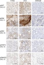
          Phosphoproteins are the key indicators of signaling network pathway activation. Many disease treatment therapies are designed to inhibit these pathways and effective diagnostics are required to evaluate the efficacy of these treatments. Phosphoprotein IHC have been impractical for diagnostics due to inconsistent results occurring from technical limitations. We have designed and tested a novel cold transport device and rapid cold plus warm formalin fixation protocol using phosphoproteins IHC. We collected 50 liver tumors that were split into two experimental conditions: 2 + 2 rapid fixation (2 hours cold then 2 hour warm formalin) or 4 hour room-temperature formalin. We analyzed primary hepatocellular carcinoma (n = 10) and metastatic gastrointestinal tumors (n = 28) for phosphoprotein IHC markers pAKT, pERK, pSRC, pSTAT3, and pSMAD2 and compared them to slides obtained from the clinical blocks. Expression of pERK and pSRC, present in the metastatic colorectal carcinoma, were better preserved with the rapid processing protocol while pSTAT3 expression was detected in hepatocellular carcinoma. Differences in pSMAD2 expression were difficult to detect due to the ubiquitous nature of protein expression. There were only 3 cases expressing pAKT and all exhibited a dramatic loss of signal for the standard clinical workflow. The rapid cold preservation shows improvement in phosphoprotein preservation.

          Related collections

          Most cited references9

          • Record: found
          • Abstract: found
          • Article: not found

          Intratumor heterogeneity in hepatocellular carcinoma.

          Morphologic intratumor heterogeneity is well known to exist in hepatocellular carcinoma (HCC), but very few systematic analyses of this phenomenon have been performed. The aim of this study was to comprehensively characterize morphologic intratumor heterogeneity in HCC. Also, taken into account were well-known immunohistochemical markers and molecular changes in liver cells that are considered in proposed classifications of liver cell neoplasms or discussed as molecular therapeutic targets.
            Bookmark
            • Record: found
            • Abstract: found
            • Article: not found

            Quantitative assessment of effect of preanalytic cold ischemic time on protein expression in breast cancer tissues.

            Companion diagnostic tests can depend on accurate measurement of protein expression in tissues. Preanalytic variables, especially cold ischemic time (time from tissue removal to fixation in formalin) can affect the measurement and may cause false-negative results. We examined 23 proteins, including four commonly used breast cancer biomarker proteins, to quantify their sensitivity to cold ischemia in breast cancer tissues. A series of 93 breast cancer specimens with known time-to-fixation represented in a tissue microarray and a second series of 25 matched pairs of core needle biopsies and breast cancer resections were used to evaluate changes in antigenicity as a function of cold ischemic time. Estrogen receptor (ER), progesterone receptor (PgR), HER2 or Ki67, and 19 other antigens were tested. Each antigen was measured using the AQUA method of quantitative immunofluorescence on at least one series. All statistical tests were two-sided. We found no evidence for loss of antigenicity with time-to-fixation for ER, PgR, HER2, or Ki67 in a 4-hour time window. However, with a bootstrapping analysis, we observed a trend toward loss for ER and PgR, a statistically significant loss of antigenicity for phosphorylated tyrosine (P = .0048), and trends toward loss for other proteins. There was evidence of increased antigenicity in acetylated lysine, AKAP13 (P = .009), and HIF1A (P = .046), which are proteins known to be expressed in conditions of hypoxia. The loss of antigenicity for phosphorylated tyrosine and increase in expression of AKAP13, and HIF1A were confirmed in the biopsy/resection series. Key breast cancer biomarkers show no evidence of loss of antigenicity, although this dataset assesses the relatively short time beyond the 1-hour limit in recent guidelines. Other proteins show changes in antigenicity in both directions. Future studies that extend the time range and normalize for heterogeneity will provide more comprehensive information on preanalytic variation due to cold ischemic time.
              Bookmark
              • Record: found
              • Abstract: found
              • Article: found
              Is Open Access

              Formalin Fixation at Low Temperature Better Preserves Nucleic Acid Integrity

              Fixation with formalin, a widely adopted procedure to preserve tissue samples, leads to extensive degradation of nucleic acids and thereby compromises procedures like microarray-based gene expression profiling. We hypothesized that RNA fragmentation is caused by activation of RNAses during the interval between formalin penetration and tissue fixation. To prevent RNAse activation, a series of tissue samples were kept under-vacuum at 4°C until fixation and then fixed at 4°C, for 24 hours, in formalin followed by 4 hours in ethanol 95%. This cold-fixation (CF) procedure preserved DNA and RNA, so that RNA segments up to 660 bp were efficiently amplified. Histological and immunohistochemical features were fully comparable with those of standard fixation. Microarray-based gene expression profiles were comparable with those obtained on matched frozen samples for probes hybridizing within 700 bases from the reverse transcription start site. In conclusion, CF preserves tissues and nucleic acids, enabling reliable gene expression profiling of fixed tissues.
                Bookmark

                Author and article information

                Contributors
                gbaird@uw.edu
                Journal
                Sci Rep
                Sci Rep
                Scientific Reports
                Nature Publishing Group UK (London )
                2045-2322
                7 February 2020
                7 February 2020
                2020
                : 10
                : 2147
                Affiliations
                [1 ]ISNI 0000 0000 8535 6057, GRID grid.412623.0, Department of Laboratory Medicine, , University of Washington Medical Center, ; Seattle, WA USA
                [2 ]ISNI 0000 0000 8535 6057, GRID grid.412623.0, Department of Surgery, , University of Washington Medical Center, ; Seattle, WA USA
                [3 ]ISNI 0000 0004 0534 4718, GRID grid.418158.1, Ventana Medical Systems, Inc., 1910 Innovation Parkway, ; Tucson, AZ USA
                [4 ]ISNI 0000000086837370, GRID grid.214458.e, Department of Pathology, , University of Michigan, ; 2800 Plymouth road, Ann Arbor, 48109 MIchigan USA
                Author information
                http://orcid.org/0000-0002-6077-0563
                http://orcid.org/0000-0001-9064-6558
                Article
                58257
                10.1038/s41598-020-58257-3
                7005752
                32034185
                a123d0a0-6e65-40d8-87c9-5e47ba0f2ddb
                © The Author(s) 2020

                Open Access This article is licensed under a Creative Commons Attribution 4.0 International License, which permits use, sharing, adaptation, distribution and reproduction in any medium or format, as long as you give appropriate credit to the original author(s) and the source, provide a link to the Creative Commons license, and indicate if changes were made. The images or other third party material in this article are included in the article’s Creative Commons license, unless indicated otherwise in a credit line to the material. If material is not included in the article’s Creative Commons license and your intended use is not permitted by statutory regulation or exceeds the permitted use, you will need to obtain permission directly from the copyright holder. To view a copy of this license, visit http://creativecommons.org/licenses/by/4.0/.

                History
                : 11 September 2019
                : 13 January 2020
                Funding
                Funded by: Ventana Medical Systems
                Categories
                Article
                Custom metadata
                © The Author(s) 2020

                Uncategorized
                liver cancer,tumour biomarkers,diagnostic markers
                Uncategorized
                liver cancer, tumour biomarkers, diagnostic markers

                Comments

                Comment on this article