1
views
0
recommends
+1 Recommend
0 collections
    0
    shares
      • Record: found
      • Abstract: found
      • Article: found
      Is Open Access

      Altered spontaneous brain activities in maintenance hemodialysis patients with cognitive impairment and the construction of cognitive function prediction models

      research-article

      Read this article at

      Bookmark
          There is no author summary for this article yet. Authors can add summaries to their articles on ScienceOpen to make them more accessible to a non-specialist audience.

          Abstract

          Objective

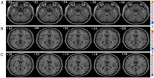
          The brain neuromechanism in maintenance hemodialysis patients (MHD) with cognitive impairment (CI) remains unclear. The study aimed to probe the relationship between spontaneous brain activity and CI by using resting-state functional magnetic resonance imaging (rs-fMRI) data.

          Methods

          Here, 55 MHD patients with CI and 28 healthy controls were recruited. For baseline data, qualitative data were compared between groups using the χ 2 test; quantitative data were compared between groups using the independent samples t-test, ANOVA test, Mann–Whitney U-test, or Kruskal–Wallis test. Comparisons of ALFF/fALFF/ReHo values among the three groups were calculated by using the DPABI toolbox, and then analyzing the correlation with clinical variables. p < .05 was considered a statistically significant difference. Furthermore, back propagation neural network (BPNN) was utilized to predict cognitive function.

          Results

          Compared with the MHD-NCI group, the patients with MHD-CI had more severe anemia and higher urea nitrogen levels, lower mALFF values in the left postcentral gyrus, lower mfALFF values in the left inferior temporal gyrus, and greater mALFF values in the right caudate nucleus ( p < .05). The above-altered indicators were correlated with MOCA scores. BPNN prediction models indicated that the diagnostic efficacy of the model which inputs were hemoglobin, urea nitrogen, and mALFF value in the left central posterior gyrus was optimal ( R 2 = 0.8054), validation cohort ( R 2 = 0.7328).

          Conclusion

          The rs-fMRI can reveal the neurophysiological mechanism of cognitive impairment in MHD patients. In addition, it can serve as a neuroimaging marker for diagnosing and evaluating cognitive impairment in MHD patients.

          Related collections

          Most cited references36

          • Record: found
          • Abstract: found
          • Article: not found

          The reward circuit: linking primate anatomy and human imaging.

          Although cells in many brain regions respond to reward, the cortical-basal ganglia circuit is at the heart of the reward system. The key structures in this network are the anterior cingulate cortex, the orbital prefrontal cortex, the ventral striatum, the ventral pallidum, and the midbrain dopamine neurons. In addition, other structures, including the dorsal prefrontal cortex, amygdala, hippocampus, thalamus, and lateral habenular nucleus, and specific brainstem structures such as the pedunculopontine nucleus, and the raphe nucleus, are key components in regulating the reward circuit. Connectivity between these areas forms a complex neural network that mediates different aspects of reward processing. Advances in neuroimaging techniques allow better spatial and temporal resolution. These studies now demonstrate that human functional and structural imaging results map increasingly close to primate anatomy.
            Bookmark
            • Record: found
            • Abstract: found
            • Article: not found

            An improved approach to detection of amplitude of low-frequency fluctuation (ALFF) for resting-state fMRI: fractional ALFF.

            Most of the resting-state functional magnetic resonance imaging (fMRI) studies demonstrated the correlations between spatially distinct brain areas from the perspective of functional connectivity or functional integration. The functional connectivity approaches do not directly provide information of the amplitude of brain activity of each brain region within a network. Alternatively, an index named amplitude of low-frequency fluctuation (ALFF) of the resting-state fMRI signal has been suggested to reflect the intensity of regional spontaneous brain activity. However, it has been indicated that the ALFF is also sensitive to the physiological noise. The current study proposed a fractional ALFF (fALFF) approach, i.e., the ratio of power spectrum of low-frequency (0.01-0.08 Hz) to that of the entire frequency range and this approach was tested in two groups of resting-state fMRI data. The results showed that the brain areas within the default mode network including posterior cingulate cortex, precuneus, medial prefrontal cortex and bilateral inferior parietal lobule had significantly higher fALFF than the other brain areas. This pattern was consistent with previous neuroimaging results. The non-specific signal components in the cistern areas in resting-state fMRI were significantly suppressed, indicating that the fALFF approach improved the sensitivity and specificity in detecting spontaneous brain activities. Its mechanism and sensitivity to abnormal brain activity should be evaluated in the future studies.
              Bookmark
              • Record: found
              • Abstract: found
              • Article: found
              Is Open Access

              Corticostriatal circuitry

              Corticostriatal connections play a central role in developing appropriate goal-directed behaviors, including the motivation and cognition to develop appropriate actions to obtain a specific outcome. The cortex projects to the striatum topographically. Thus, different regions of the striatum have been associated with these different functions: the ventral striatum with reward; the caudate nucleus with cognition; and the putamen with motor control. However, corticostriatal connections are more complex, and interactions between functional territories are extensive. These interactions occur in specific regions in which convergence of terminal fields from different functional cortical regions are found. This article provides an overview of the connections of the cortex to the striatum and their role in integrating information across reward, cognitive, and motor functions. Emphasis is placed on the interface between functional domains within the striatum.
                Bookmark

                Author and article information

                Journal
                Ren Fail
                Ren Fail
                Renal Failure
                Taylor & Francis
                0886-022X
                1525-6049
                29 May 2023
                2023
                29 May 2023
                : 45
                : 1
                : 2217276
                Affiliations
                [a ]Department of Nephrology, The Affiliated Changzhou NO.2 People’s Hospital of Nanjing Medical University , Changzhou, Jiangsu, China
                [b ]Graduate College, Dalian Medical University , Dalian, China
                [c ]Department of Radiology, The Affiliated Changzhou NO.2 People’s Hospital of Nanjing Medical University , Changzhou, Jiangsu, China
                [d ]School of Microelectronics and Control Engineering, Changzhou University , Changzhou, China
                [e ]School of Computer Science and Artificial Intelligence, Changzhou University , Changzhou, China
                [f ]Hemodialysis Center, The Affiliated Changzhou NO.2 People’s Hospital of Nanjing Medical University , Changzhou, Jiangsu, China
                Author notes
                [*]

                Qing Sun and Jiahui Zheng contributed equally to this work.

                CONTACT Tongqiang Liu liuyf1106@ 123456126.com Department of Nephrology, The Affiliated Changzhou NO.2 People’s Hospital of Nanjing Medical University , Changzhou, Jiangsu, China
                Haifeng Shi 18114330152@ 123456163.com Department of Radiology, The Affiliated Changzhou NO.2 People’s Hospital of Nanjing Medical University , Changzhou, Jiangsu, China
                Author information
                https://orcid.org/0000-0002-5990-7251
                Article
                2217276
                10.1080/0886022X.2023.2217276
                10228305
                37246750
                9f74558a-2492-4e9f-9337-d4ce96610ffa
                © 2023 The Author(s). Published by Informa UK Limited, trading as Taylor & Francis Group.

                This is an Open Access article distributed under the terms of the Creative Commons Attribution-NonCommercial License ( http://creativecommons.org/licenses/by-nc/4.0/), which permits unrestricted non-commercial use, distribution, and reproduction in any medium, provided the original work is properly cited. The terms on which this article has been published allow the posting of the Accepted Manuscript in a repository by the author(s) or with their consent.

                History
                Page count
                Figures: 7, Tables: 6, Pages: 12, Words: 7258
                Categories
                Research Article
                Research Article
                Clinical Study

                Nephrology
                resting-state magnetic resonance imaging,maintenance hemodialysis,cognitive impairment,low-frequency amplitude,ratiometric low-frequency amplitude,regional homogeneity,back propagation neural network

                Comments

                Comment on this article