0
views
0
recommends
+1 Recommend
0 collections
    0
    shares
      • Record: found
      • Abstract: found
      • Article: found
      Is Open Access

      Variations in the Size and Shape of Human Cochlear Malformation Types

      research-article

      Read this article at

      Bookmark
          There is no author summary for this article yet. Authors can add summaries to their articles on ScienceOpen to make them more accessible to a non-specialist audience.

          ABSTRACT

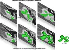
          The objective of this study is to determine the variations in size and shape of the most widely recognized cochlear malformation types using three‐dimensional (3D) visualization. Using 3D slicer freeware, the complete inner‐ear structures were segmented from 46 anonymized high‐resolution computed tomography (HRCT) image datasets. Cochlear height, internal auditory canal height, and width were measured from the axial plane. Cochlear basal turn diameter was measured from the oblique coronal plane. Number of cochlear turns was measured from the 3D images and the corresponding cochlear duct length (CDL) was estimated using the CDL equations given in Alexiades et al. [Otol Neurotol 36 (2015) 904–907]. Out of 46 preoperative HRCT image datasets of human temporal bone, cochlear anatomy types including normal anatomy (4), enlarged vestibular aqueduct syndrome (3), cochlear aplasia (2), incomplete partition Types I (8), II (Mondini's deformity) (3), and III (X‐linked) (4), cochlear hypoplasia (CH) (17), and common cavity (CC) (5) were identified. Majority of CH cases had cochlear height shorter than 4 mm whereas the CC cases measured cochlear height above 6 mm. For all the other malformation types, cochlear height was between 4 and 6 mm. In terms of “A” value, majority of CH cases showed shorter “A” value of <7.5 mm, which is in the lower end in comparison to the rest of the malformation types reported in this study. 3D‐visualization shows the size and shape variations of all the structures of inner ear and also improves the clinicians' ability to visualize cochlear anatomy and nearby structures much easier than from the 2D image slices. Anat Rec, 302:1792–1799, 2019. © 2019 The Author. The Anatomical Record published by Wiley Periodicals, Inc. on behalf of American Association for Anatomy

          Related collections

          Most cited references15

          • Record: found
          • Abstract: found
          • Article: not found

          A new classification for cochleovestibular malformations.

          The report proposes a new classification system for inner ear malformations, based on radiological features of inner ear malformations reviewed in 23 patients. The investigation took the form of a retrospective review of computerized tomography findings relating to the temporal bone in 23 patients (13 male and 10 female patients) with inner ear malformations. The subjects were patients with profound bilateral sensorineural hearing loss who had all had high-resolution computed tomography (CT) with contiguous 1-mm-thick images obtained through the petrous bone in axial sections. The CT results were reviewed for malformations of bony otic capsule under the following subgroups: cochlear, vestibular, semicircular canal, internal auditory canal (IAC), and vestibular and cochlear aqueduct malformations. Cochlear malformations were classified as Michel deformity, common cavity deformity, cochlear aplasia, hypoplastic cochlea, incomplete partition types I (IP-I) and II (IP-II) (Mondini deformity). Incomplete partition type I (cystic cochleovestibular malformation) is defined as a malformation in which the cochlea lacks the entire modiolus and cribriform area, resulting in a cystic appearance, and there is an accompanying large cystic vestibule. In IP-II (the Mondini deformity), there is a cochlea consisting of 1.5 turns (in which the middle and apical turns coalesce to form a cystic apex) accompanied by a dilated vestibule and enlarged vestibular aqueduct. Four patients demonstrated anomalies involving only one inner ear component. All the remaining patients had diseases or conditions affecting more than one inner ear component. Eight ears had IP-I, and 10 patients had IP-II. Ears with IP-I had large cystic vestibules, whereas the amount of dilation was minimal in patients with IP-II. The majority of the semicircular canals (67%) were normal. Semicircular canal aplasia accompanied cases of Michel deformity, cochlear hypoplasia, and common cavity. In 14 ears, the IAC had a defective fundus at the lateral end. In two ears the IAC was absent. In all seven cases of common cavity malformations, there was a bony defect at the lateral end of the IAC. In five of them the IAC was enlarged, whereas in two the IAC was narrow. All patients with IP-I had an enlarged IAC, whereas in patients with type II disease, four had a normal IAC and 10 had an enlarged IAC. All cases of IP-II had an enlarged vestibular aqueduct, whereas this finding was not present in any of the cases of IP-I. In all cases, the vestibular aqueduct findings were symmetrical on both sides (simultaneously normal or enlarged). No patient demonstrated enlargement or any other abnormalities involving the cochlear aqueduct. Radiological findings of congenital malformations in the present study suggested two different types of incomplete partition. Cystic cochleovestibular malformation (IP-I) and the classic Mondini deformity (IP-II). The type I malformation is less differentiated than the type II malformation. Classic Mondini deformity has three components (a cystic apex, dilated vestibule, and large vestibular aqueduct), whereas type I malformation has an empty, cystic cochlea and vestibule without an enlarged vestibular aqueduct. Mondini deformity represents a later malformation, so the amount of dysplasia is much less than in type II. Therefore, it is more accurate and useful for clinical purposes to classify these malformations (in descending order of severity) as follows: Michel deformity, cochlear aplasia, common cavity, IP-I (cystic cochleovestibular malformation), cochlear hypoplasia, and IP-II (Mondini deformity). Only in this way can these complex malformations be grouped precisely and the results of cochlear implantation compared.
            Bookmark
            • Record: found
            • Abstract: found
            • Article: not found
            Is Open Access

            An overview of cochlear implant electrode array designs

            Cochlear implant electrode arrays are designed with specific characteristics that allow for the preservation of intra-cochlear structures during the insertion process, as well as during explantation. Straight lateral wall (LW) electrode arrays and pre-curved modiolar hugging (MH) electrode arrays are the two types that are commercially available. Although there is a third type of electrode array called the mid-scala (MS), which is positioned in the middle of the scala tympani (ST), and is usually considered as an MH type of electrode. Different lengths of straight LW electrode arrays are currently available which allow for insertion across a range of different sized cochleae; however, due to manufacturing limitations, pre-curved MH electrodes are generally only available to cover the basal turn of the cochlea, while the spiral ganglion cells are distributed in the Rosenthal's canal that extends into 1.75 turns of the cochlea. Both straight LW and pre-curved MH electrodes can cause a certain degree of intra-cochlear trauma, but pre-curved MH electrodes tend to deviate into the scala vestibuli from the scala tympani more often than the straight LW electrodes, resulting in damage to the osseous spiral lamina/spiral ligament which could initiate new bone formation and eventually affect the cochlear implant users' hearing performance. Structural damage to the cochlea could also affect the vestibular function. With pre-curved MH electrodes, higher degrees of trauma are related to the fixed curling geometry of the electrode in relation to the variable coiling pattern of individual cochleae, the orientation of the electrode contacts in relation to the modiolus wall, and how effectively the stylet was handled by the surgeon during the procedure. Wire management, metal density, and the shore hardness of the silicone elastomer all contribute to the stiffness/flexibility of the electrode. It is important to acknowledge the impact of bringing the stimulating contacts closer to the modiolus wall with an MH electrode type in terms of the resultant damage to intra-cochlear structures. The presence of malformed cochleae should be identified and appropriate electrodes should be chosen for each specific cochlea, irrespective of the cochlear implant brand. In order to utilize drug therapy, the cochlea should be free from any trauma.
              Bookmark
              • Record: found
              • Abstract: found
              • Article: found
              Is Open Access

              Classification and Current Management of Inner Ear Malformations

              Morphologically congenital sensorineural hearing loss can be investigated under two categories. The majority of congenital hearing loss causes (80%) are membranous malformations. Here, the pathology involves inner ear hair cells. There is no gross bony abnormality and, therefore, in these cases high-resolution computerized tomography and magnetic resonance imaging of the temporal bone reveal normal findings. The remaining 20% have various malformations involving the bony labyrinth and, therefore, can be radiologically demonstrated by computerized tomography and magnetic resonance imaging. The latter group involves surgical challenges as well as problems in decision-making. Some cases may be managed by a hearing aid, others need cochlear implantation, and some cases are candidates for an auditory brainstem implantation (ABI). During cochlear implantation, there may be facial nerve abnormalities, cerebrospinal fluid leakage, electrode misplacement or difficulty in finding the cochlea itself. During surgery for inner ear malformations, the surgeon must be ready to modify the surgical approach or choose special electrodes for surgery. In the present review article, inner ear malformations are classified according to the differences observed in the cochlea. Hearing and language outcomes after various implantation methods are closely related to the status of the cochlear nerve, and a practical classification of the cochlear nerve deficiency is also provided.
                Bookmark

                Author and article information

                Contributors
                anandhan.dhanasingh@medel.com
                Journal
                Anat Rec (Hoboken)
                Anat Rec (Hoboken)
                10.1002/(ISSN)1932-8494
                AR
                Anatomical Record (Hoboken, N.j. : 2007)
                John Wiley & Sons, Inc. (Hoboken, USA )
                1932-8486
                1932-8494
                24 April 2019
                October 2019
                : 302
                : 10 ( doiID: 10.1002/ar.v302.10 )
                : 1792-1799
                Affiliations
                [ 1 ] MED‐EL GmbH Innsbruck Austria
                Author notes
                [*] [* ] Correspondence to: Anandhan Dhanasingh, MED‐EL GmbH, Fürstenweg 77a, Innsbruck 6020, Austria E‐mail: anandhan.dhanasingh@ 123456medel.com

                Author information
                https://orcid.org/0000-0003-2116-9318
                Article
                AR24136
                10.1002/ar.24136
                6767414
                30980504
                9dea9b1b-bcc4-43a8-936f-4034066a6fcf
                © 2019 The Author. The Anatomical Record published by Wiley Periodicals, Inc. on behalf of American Association for Anatomy

                This is an open access article under the terms of the http://creativecommons.org/licenses/by/4.0/ License, which permits use, distribution and reproduction in any medium, provided the original work is properly cited.

                History
                : 16 October 2018
                : 08 January 2019
                : 28 January 2019
                Page count
                Figures: 6, Tables: 2, Pages: 8, Words: 3221
                Categories
                Hearing Biology
                Hearing Biology
                Custom metadata
                2.0
                ar24136
                October 2019
                Converter:WILEY_ML3GV2_TO_NLMPMC version:5.6.9 mode:remove_FC converted:30.09.2019

                Anatomy & Physiology
                inner ear malformation,3d visualization,size and shape variation,cochlear height,basal turn diameter

                Comments

                Comment on this article