10
views
0
recommends
+1 Recommend
0 collections
    0
    shares
      • Record: found
      • Abstract: found
      • Article: found
      Is Open Access

      Propofol inhibits oxidative stress injury through the glycogen synthase kinase 3 beta/nuclear factor erythroid 2-related factor 2/heme oxygenase-1 signaling pathway

      research-article
      a , b , c , d , c
      Bioengineered
      Taylor & Francis
      Propofol, cardiomyocyte injury, oxidative stress, myocardial ischemic reperfusion injury (MIRI), GSK3β-Nrf2/HO-1

      Read this article at

      Bookmark
          There is no author summary for this article yet. Authors can add summaries to their articles on ScienceOpen to make them more accessible to a non-specialist audience.

          ABSTRACT

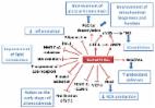
          Oxidative stress is the main cause of ischemia/reperfusion injury. Propofol is a commonly used intravenous hypnotic anesthetic agent with antioxidant properties. In this study, we aimed to elucidate the protective effects of propofol on H 2O 2-induced cardiomyocyte injury and myocardial ischemic/reperfusion injury (MIRI) in rats. Cardiomyocyte injury was evaluated by determining cardiac troponin-1 (cTn-1) and creatine kinase-MB (CK-MB) levels. Antioxidative stress was assessed by measuring lactate dehydrogenase (LDH), malondialdehyde (MDA), glutathione (GSH), superoxide dismutase (SOD), reactive oxygen species (ROS), and catalase (CAT) levels. Apoptosis was evaluated using flow cytometry and TUNEL assays. Bax and Bcl-2 expression levels were determined by quantitative reverse transcription PCR (qRT-PCR) and Western blotting. The levels of glycogen synthase kinase 3 beta/nuclear factor erythroid 2-related factor 2 (Nrf2)/heme oxygenase-1 (HO-1) pathway-related factors were measured using Western blotting. Myocardial infarction in rats was analyzed using an Evans blue staining assay. The results showed that propofol reduced the levels of CK-MB, cTn-1, LDH, MDA, and ROS, and increased the levels of GSH, SOD, and CAT in H 2O 2-treated H9c2 cells. Additionally, propofol inhibited H 2O 2-induced apoptosis by downregulating Bax and upregulating Bcl-2. Moreover, propofol decreased the area of myocardial infarction in rats with MIRI. The GSK3β-Nrf2/HO-1 signaling pathway was activated by propofol. Rescue experiments showed that Nrf2 knockdown alleviated the effects of propofol on oxidative stress and apoptosis in H9c2 cells. In conclusion, propofol attenuated H 2O 2-induced myocardial cell injury by regulating the GSK3β/Nrf2/HO-1 signaling pathway and alleviating MIRI, suggesting that propofol is a promising therapeutic option for ischemic heart disease.

          Related collections

          Most cited references43

          • Record: found
          • Abstract: found
          • Article: not found

          Nrf2, a Cap'n'Collar transcription factor, regulates induction of the heme oxygenase-1 gene.

          Stress response elements, which mediate induction of the mouse heme oxygenase-1 (HO-1) gene by several agents, resemble the binding site for the activator protein-1 (Jun/Fos), Maf, and Cap'n'Collar/basic leucine zipper (CNC-bZIP) families of proteins. In L929 fibroblasts, significant activation of an HO-1 enhancer-reporter fusion gene was observed only with the CNC-bZIP class of proteins with Nrf2 exhibiting the highest level of trans-activation, between 25- and 30-fold. To further examine the role of this factor in HO-1 gene regulation, a dominant-negative mutant, Nrf2M, was generated and conditionally expressed in L929 cells. The mutant protein was detected in cytoplasmic and nuclear fractions but did not affect cell growth. Under conditions of Nrf2M overexpression, HO-1 mRNA accumulation in response to heme, cadmium, zinc, arsenite, and tert-butylhydroquinone was inhibited by 85-95%. In contrast, overexpression of a dominant-negative mutant of c-Jun decreased L929 cell growth but did not inhibit HO-1 gene activation. Nrf2 does not homodimerize, but CNC-bZIP.small Maf protein heterodimers and Nrf2. Jun protein complexes are proposed to function as trans-activators. Co-expression of Jun proteins or p18, however, had no significant affect or inhibited Nrf2-mediated trans-activation. Taken together, these results implicate Nrf2 in the induction of the HO-1 gene but suggest that the Nrf2 partner in this function is a factor other than p18 or Jun proteins.
            Bookmark
            • Record: found
            • Abstract: found
            • Article: found
            Is Open Access

            Resveratrol and Cardiovascular Diseases

            The increased incidence of cardiovascular diseases (CVDs) has stimulated research for substances that could improve cardiovascular health. Among them, resveratrol (RES), a polyphenolic compound notably present in grapes and red wine, has been involved in the “French paradox”. RES is known for its antioxidant and anti-inflammatory properties and for its ability to upregulate endothelial NO synthase (eNOS). RES was able to scavenge •OH/O2 •− and peroxyl radicals, which can limit the lipid peroxidation processes. Moreover, in bovine aortic endothelial cells (BAEC) under glucose-induced oxidative stress, RES restored the activity of dimethylargininedimethylaminohydrolase (DDAH), an enzyme that degrades an endogenous inhibitor of eNOS named asymmetric dimethylarginine (ADMA). Thus, RES could improve •NO availability and decrease the endothelial dysfunction observed in diabetes. Preclinical studies have made it possible to identify molecular targets (SIRT-1, AMPK, Nrf2, NFκB…); however, there are limited human clinical trials, and difficulties in the interpretation of results arise from the use of high-dose RES supplements in research studies, whereas low RES concentrations are present in red wine. The discussions on potential beneficial effects of RES in CVDs (atherosclerosis, hypertension, stroke, myocardial infarction, heart failure) should compare the results of preclinical studies with those of clinical trials.
              Bookmark
              • Record: found
              • Abstract: found
              • Article: not found

              Pitx2 promotes heart repair by activating the antioxidant response after cardiac injury

              Summary Myocardial infarction results in compromised myocardial function with heart failure due to insufficient cardiomyocyte self-renewal 1 . Unlike lower vertebrates, mammalian hearts only have a transient neonatal renewal capacity 2 . Reactivating primitive reparative ability in the mature heart requires knowledge of the mechanisms promoting early heart repair. By testing an established Hippo-deficient heart regeneration model for renewal promoting factors, we found that Pitx2 expression was induced in injured, Hippo-deficient ventricles. Pitx2-deficient neonatal hearts failed to repair after apex resection while Pitx2-gain-of-function in adult cardiomyocytes conferred reparative ability after myocardial infarction. Genomic analyses indicated that Pitx2 activated genes encoding electron transport chain components and reactive oxygen species scavengers. A subset of Pitx2 target genes was cooperatively regulated with the Hippo effector, Yap. Furthermore, Nrf2, a regulator of antioxidant response 3 , directly regulated Pitx2 expression and subcellular localization. Pitx2 mutant myocardium had elevated reactive oxygen species levels while antioxidant supplementation suppressed the Pitx2-loss-of-function phenotype. These findings reveal a genetic pathway, activated by tissue damage that is essential for cardiac repair.
                Bookmark

                Author and article information

                Journal
                Bioengineered
                Bioengineered
                Bioengineered
                Taylor & Francis
                2165-5979
                2165-5987
                14 January 2022
                2022
                14 January 2022
                : 13
                : 1
                : 1612-1625
                Affiliations
                [a ]Department of Anesthesiology, The First Affiliated Hospital of Guangzhou University of Chinese Medicine; , Guangdong, China
                [b ]Department of Anesthesiology, Fifth Medical Center of Chinese Pla General Hospital; , Beijing, China
                [c ]Department of Anesthesiology, The Second Affiliated Hospital of Guangzhou University of Chinese Medicine; , Guangdong, China
                [d ]Department of Medical Administration, The Eleventh People’s Hospital of Guangzhou; , Guangdong, China
                Author notes
                CONTACT Jinfeng Li ljf2525@ 123456163.com The Second Affiliated Hospital of Guangzhou University of Chinese Medicine; , No. 261 Datong Road, Yuexiu District, Guangzhou, Guangdong 510105, China
                [#]

                Ziyin Zhang and Baofeng Yan contributed equally in this work.

                Article
                2021062
                10.1080/21655979.2021.2021062
                8805835
                35030972
                9bba972a-5108-46b9-95c0-b5d331ee07d3
                © 2022 The Author(s). Published by Informa UK Limited, trading as Taylor & Francis Group.

                This is an Open Access article distributed under the terms of the Creative Commons Attribution License ( http://creativecommons.org/licenses/by/4.0/), which permits unrestricted use, distribution, and reproduction in any medium, provided the original work is properly cited.

                History
                Page count
                Figures: 10, References: 43, Pages: 14
                Categories
                Research Article
                Research Paper

                Biomedical engineering
                propofol,cardiomyocyte injury,oxidative stress,myocardial ischemic reperfusion injury (miri),gsk3β-nrf2/ho-1

                Comments

                Comment on this article