1
views
0
recommends
+1 Recommend
0 collections
    0
    shares
      • Record: found
      • Abstract: found
      • Article: found
      Is Open Access

      Ex vivo, in situ perfusion protocol for human brain fixation compatible with microscopy, MRI techniques, and anatomical studies

      methods-article
      1 , * , , 2 , 1 , 1 , 1 , 1 , 1 , 1 , 1 , 1 , 1 , 1 , 1 , 3 , 3 , 4 , 5 , 6 , 3 , 5 , 1 , * ,
      Frontiers in Neuroanatomy
      Frontiers Media S.A.
      human brain, fixation, carotid perfusion, histology, electron microscopy, intracellular injection, MRI

      Read this article at

      Bookmark
          There is no author summary for this article yet. Authors can add summaries to their articles on ScienceOpen to make them more accessible to a non-specialist audience.

          Abstract

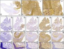
          We present a method for human brain fixation based on simultaneous perfusion of 4% paraformaldehyde through carotids after a flush with saline. The left carotid cannula is used to perfuse the body with 10% formalin, to allow further use of the body for anatomical research or teaching. The aim of our method is to develop a vascular fixation protocol for the human brain, by adapting protocols that are commonly used in experimental animal studies. We show that a variety of histological procedures can be carried out (cyto- and myeloarchitectonics, histochemistry, immunohistochemistry, intracellular cell injection, and electron microscopy). In addition, ex vivo, ex situ high-resolution MRI (9.4T) can be obtained in the same specimens. This procedure resulted in similar morphological features to those obtained by intravascular perfusion in experimental animals, provided that the postmortem interval was under 10 h for several of the techniques used and under 4 h in the case of intracellular injections and electron microscopy. The use of intravascular fixation of the brain inside the skull provides a fixed whole human brain, perfectly fitted to the skull, with negligible deformation compared to conventional techniques. Given this characteristic of ex vivo, in situ fixation, this procedure can probably be considered the most suitable one available for ex vivo MRI scans of the brain. We describe the compatibility of the method proposed for intravascular fixation of the human brain and fixation of the donor’s body for anatomical purposes. Thus, body donor programs can provide human brain tissue, while the remainder of the body can also be fixed for anatomical studies. Therefore, this method of human brain fixation through the carotid system optimizes the procurement of human brain tissue, allowing a greater understanding of human neurological diseases, while benefiting anatomy departments by making the remainder of the body available for teaching purposes.

          Related collections

          Most cited references82

          • Record: found
          • Abstract: found
          • Article: found
          Is Open Access

          A probabilistic atlas of the human thalamic nuclei combining ex vivo MRI and histology

          The human thalamus is a brain structure that comprises numerous, highly specific nuclei. Since these nuclei are known to have different functions and to be connected to different areas of the cerebral cortex, it is of great interest for the neuroimaging community to study their volume, shape and connectivity in vivo with MRI. In this study, we present a probabilistic atlas of the thalamic nuclei built using ex vivo brain MRI scans and histological data, as well as the application of the atlas to in vivo MRI segmentation. The atlas was built using manual delineation of 26 thalamic nuclei on the serial histology of 12 whole thalami from six autopsy samples, combined with manual segmentations of the whole thalamus and surrounding structures (caudate, putamen, hippocampus, etc.) made on in vivo brain MR data from 39 subjects. The 3D structure of the histological data and corresponding manual segmentations was recovered using the ex vivo MRI as reference frame, and stacks of blockface photographs acquired during the sectioning as intermediate target. The atlas, which was encoded as an adaptive tetrahedral mesh, shows a good agreement with previous histological studies of the thalamus in terms of volumes of representative nuclei. When applied to segmentation of in vivo scans using Bayesian inference, the atlas shows excellent test-retest reliability, robustness to changes in input MRI contrast, and ability to detect differential thalamic effects in subjects with Alzheimer's disease. The probabilistic atlas and companion segmentation tool are publicly available as part of the neuroimaging package FreeSurfer.
            Bookmark
            • Record: found
            • Abstract: found
            • Article: not found

            Quantitative comparison of 21 protocols for labeling hippocampal subfields and parahippocampal subregions in in vivo MRI: towards a harmonized segmentation protocol.

            An increasing number of human in vivo magnetic resonance imaging (MRI) studies have focused on examining the structure and function of the subfields of the hippocampal formation (the dentate gyrus, CA fields 1-3, and the subiculum) and subregions of the parahippocampal gyrus (entorhinal, perirhinal, and parahippocampal cortices). The ability to interpret the results of such studies and to relate them to each other would be improved if a common standard existed for labeling hippocampal subfields and parahippocampal subregions. Currently, research groups label different subsets of structures and use different rules, landmarks, and cues to define their anatomical extents. This paper characterizes, both qualitatively and quantitatively, the variability in the existing manual segmentation protocols for labeling hippocampal and parahippocampal substructures in MRI, with the goal of guiding subsequent work on developing a harmonized substructure segmentation protocol.
              Bookmark
              • Record: found
              • Abstract: found
              • Article: not found

              MR volumetric analysis of the human entorhinal, perirhinal, and temporopolar cortices.

              Our purpose was to investigate the normal volumes of the human entorhinal, perirhinal, and temporopolar cortices on MR imaging studies using a customized program. We designed a protocol in which the volumes of the entorhinal, perirhinal, and temporopolar cortices were determined from coronal MR images using anatomic landmarks defined on the basis of cytoarchitectonic analyses of 49 autopsy cases. MR volumetry of these cortical areas was performed in 52 healthy volunteers. The overall mean volumes were 1768 +/- 328 mm3/1558 +/- 341 mm3 (right/left) for the entorhinal cortex, 2512 +/- 672 mm3/2572 +/- 666 mm3 for the perirhinal cortex, and 2960 +/- 623 mm3/3091 +/- 636 mm3 for the temporopolar cortex. The right entorhinal cortex was 12% larger than the left. The volume of the temporopolar cortex was reduced bilaterally by 13% in the older age group compared with younger subjects, while the volumes of the entorhinal and perirhinal cortices were unaffected by age. There were no differences between men and women in the volumes of any of the three cortices. Our method provides a tool by which to measure volumes of the entorhinal, perirhinal, and temporopolar cortices on coronal MR images.
                Bookmark

                Author and article information

                Contributors
                Journal
                Front Neuroanat
                Front Neuroanat
                Front. Neuroanat.
                Frontiers in Neuroanatomy
                Frontiers Media S.A.
                1662-5129
                23 March 2023
                2023
                : 17
                : 1149674
                Affiliations
                [1] 1Human Neuroanatomy Laboratory, Neuromax CSIC Associated Unit, Medical Sciences Department, School of Medicine and CRIB, University of Castilla La Mancha , Albacete, Spain
                [2] 2Department of Health, School of Medicine, Public University of Navarra , Pamplona, Spain
                [3] 3Laboratorio Cajal de Circuitos Corticales, Centro de Tecnología Biomédica, Universidad Politécnica de Madrid, and Instituto Cajal, CSIC , Madrid, Spain
                [4] 4Department of Diagnostic Radiology, Lund University , Lund, Sweden
                [5] 5Department of Radiology, University of Pennsylvania , Philadelphia, PA, United States
                [6] 6Department of Neurology, University of Pennsylvania , Philadelphia, PA, United States
                Author notes

                Edited by: Basilis Zikopoulos, Boston University, United States

                Reviewed by: Miguel Ángel García-Cabezas, Autonomous University of Madrid, Spain; Alejandra Sierra, University of Eastern Finland, Finland

                *Correspondence: Ricardo Insausti ricardo.insausti@ 123456uclm.es Emilio Artacho-Pérula emilio.artacho@ 123456uclm.es
                Article
                10.3389/fnana.2023.1149674
                10076536
                37034833
                9b6f5d0f-6d72-4f69-a5c7-01439da73e73
                Copyright © 2023 Insausti, Insausti, Muñoz López, Medina Lorenzo, Arroyo-Jiménez, Marcos Rabal, de La Rosa-Prieto, Delgado-González, Montón Etxeberria, Cebada-Sánchez, Raspeño-García, Iñiguez de Onzoño, Molina Romero, Benavides-Piccione, Tapia-González, Wisse, Ravikumar, Wolk, DeFelipe, Yushkevich and Artacho-Pérula.

                This is an open-access article distributed under the terms of the Creative Commons Attribution License (CC BY). The use, distribution or reproduction in other forums is permitted, provided the original author(s) and the copyright owner(s) are credited and that the original publication in this journal is cited, in accordance with accepted academic practice. No use, distribution or reproduction is permitted which does not comply with these terms.

                History
                : 22 January 2023
                : 28 February 2023
                Page count
                Figures: 13, Tables: 1, Equations: 0, References: 82, Pages: 23, Words: 182065
                Funding
                Funded by: National Institutes of Health, doi 10.13039/100000002;
                Award ID: R01 AG056014-R01
                This work was supported in part by the National Institute of Health (Grant R01 AG056014-R01), a UCLM travel grant (to RI), a UCLM research grant to Human Neuroanatomy Laboratory (2020-GRIN-28837), and project EQC2019-006341-P (AEI/FEDER UE).
                Categories
                Neuroscience
                Methods

                Neurosciences
                human brain,fixation,carotid perfusion,histology,electron microscopy,intracellular injection,mri

                Comments

                Comment on this article