9
views
0
recommends
+1 Recommend
0 collections
    0
    shares
      • Record: found
      • Abstract: found
      • Article: found
      Is Open Access

      Correlation between central stromal demarcation line depth and changes in K values after corneal cross-linking (CXL)

      research-article

      Read this article at

      Bookmark
          There is no author summary for this article yet. Authors can add summaries to their articles on ScienceOpen to make them more accessible to a non-specialist audience.

          Abstract

          Purpose

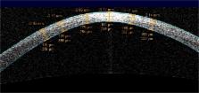
          A stromal demarcation line (DL) after corneal cross-linking (CXL) has lately been suggested as a surrogate parameter for the success of CXL. The aim of this study was to investigate the correlation between depth of the central DL 1 month and the change in K values 12 months after CXL.

          Methods

          Treatment-naive subjects with keratoconus were treated using an accelerated CXL protocol [A-CXL(9*10)]. Depth of the DL/relative depth of the DL (DL%) was measured using Visante OCT imaging 1 month postoperatively (OP). K max/ K 2.5 (preOP) and change in K max/ K 2.5 (preOP − 12 months postOP) were assessed using corneal tomography (Pentacam HR, Oculus GmBH).

          Results

          Forty eyes were treated following the A-CXL(9*10). The mean DL depth was 200 ± 99 μm (range 71 to 479)/mean DL% = 42.70 ± 20.00% (range 17–90). There was no statistically significant correlation between stromal depth of the DL and change in K max or K 2.5, respectively (Spearman rho DL/∆ K max − 0.14 and DL/∆ K 2.5 − 0.14). Between DL% and the changes in maximum K values or K 2.5, no statistically significant correlation was found as well (Spearman rho DL%/∆ K max − 0.10 and DL%/∆ K 2.5 − 0.19). Mean change in K max after 12 months was − 0.68 ± 2.26 diopters (D) (median − 0.35 D) and − 0.82 ± 1.6 D (median − 0.65 D) for K 2.5 ( p = 0.07; p = 0.02).

          Conclusions

          No statistically significant correlation was found between the stromal central depth of the DL and any outcome parameter for CXL after 12 months. Therefore, the interpretation of the DL as a predictive parameter for the effect of the procedure may not apply.

          Related collections

          Most cited references19

          • Record: found
          • Abstract: found
          • Article: not found

          Corneal cross-linking-induced stromal demarcation line.

          Corneal collagen cross-linking by UVA/riboflavin (X-linking) represents a new method for the treatment of progressive keratoconus and currently is under clinical study. To avoid UVA irradiation damage to the corneal endothelium, the parameters for X-linking are set in a way that effective treatment occurs only in the first 300 microm of the corneal stroma. Here, X-linking not only strengthens the biomechanical properties of the cornea but also induces keratocyte apoptosis. To date, the effectiveness of treatment could be monitored only indirectly by postoperative follow-up corneal topographies or using corneal confocal microscopy. Here we describe a corneal stromal demarcation line indicating the transition zone between cross-linked anterior corneal stroma and untreated posterior corneal stroma. The demarcation line is biomicroscopically detectable in slit-lamp examination as early as 2 weeks after treatment. X-linking was performed in 16 cases of progressive keratoconus, and corneas were examined biomicroscopically and by means of corneal topography and pachymetry before and after treatment. In 14 of 16 cases, a thin stromal demarcation line was visible at a depth of approximately 300 microm over the whole cornea after X-linking treatment. This newly observed demarcation line may result from differences in the refractive index and/or reflection properties of untreated versus X-linked corneal stroma and represents an effective tool to biomicroscopically easily monitor the depth of effective X-linking treatment in keratoconus.
            Bookmark
            • Record: found
            • Abstract: found
            • Article: not found

            Treatment of progressive keratoconus by riboflavin-UVA-induced cross-linking of corneal collagen: ultrastructural analysis by Heidelberg Retinal Tomograph II in vivo confocal microscopy in humans.

            To assess ultrastructural stromal modifications after riboflavin-UVA-induced cross-linking of corneal collagen in patients with progressive keratoconus. This was a second-phase prospective nonrandomized open study in 10 patients with progressive keratoconus treated by riboflavin-UVA-induced cross-linking of corneal collagen and assessed by means of Heidelberg Retinal Tomograph II Rostock Corneal Module (HRT II-RCM) in vivo confocal microscopy. The eye in the worst clinical condition was treated for each patient. Treatment under topical anesthesia included corneal deepithelization (9-mm diameter) and instillation of 0.1% riboflavin phosphate-20% dextran T 500 solution at 5 minutes before UVA irradiation and every 5 minutes for a total of 30 minutes. UVA irradiation was 7 mm in diameter. Patients were assessed by HRT II-RCM confocal microscopy in vivo at 1, 3, and 6 months after treatment. Rarefaction of keratocytes in the anterior and intermediate stroma, associated with stromal edema, was observed immediately after treatment. The observation at 3 months after the operation detected keratocyte repopulation in the central treated area, whereas the edema had disappeared. Cell density increased progressively over the postoperative period. At approximately 6 months, keratocyte repopulation was complete, accompanied by increased density of stromal fibers. No endothelial damage was observed at any time. Reduction in anterior and intermediate stromal keratocytes followed by gradual repopulation has been confirmed directly in vivo in humans by HRT II-RCM confocal microscopy after riboflavin-UVA-induced corneal collagen cross-linking.
              Bookmark
              • Record: found
              • Abstract: found
              • Article: not found

              Corneal biomechanical properties at different corneal cross-linking (CXL) irradiances.

              New corneal cross-linking (CXL) devices are capable of using higher UV-A light irradiances than used in original CXL protocols. The Bunsen-Roscoe law states that a photochemical reaction should stay constant if the delivered total energy is kept constant; however, little clinical data are available to support this hypothesis.
                Bookmark

                Author and article information

                Contributors
                gerald.schmidinger@meduniwien.ac.at
                Journal
                Graefes Arch Clin Exp Ophthalmol
                Graefes Arch. Clin. Exp. Ophthalmol
                Graefe's Archive for Clinical and Experimental Ophthalmology
                Springer Berlin Heidelberg (Berlin/Heidelberg )
                0721-832X
                1435-702X
                23 February 2018
                23 February 2018
                2018
                : 256
                : 4
                : 759-764
                Affiliations
                ISNI 0000 0000 9259 8492, GRID grid.22937.3d, Department of Ophthalmology, , Medical University of Vienna, ; Waehringer Guertel 18-20, 1090 Vienna, Austria
                Article
                3922
                10.1007/s00417-018-3922-z
                5856899
                29476249
                9a8f8656-af57-4205-a84b-09a9e0b05cab
                © The Author(s) 2018

                Open Access This article is distributed under the terms of the Creative Commons Attribution 4.0 International License (http://creativecommons.org/licenses/by/4.0/), which permits unrestricted use, distribution, and reproduction in any medium, provided you give appropriate credit to the original author(s) and the source, provide a link to the Creative Commons license, and indicate if changes were made.

                History
                : 15 September 2017
                : 8 January 2018
                : 24 January 2018
                Funding
                Funded by: Medical University of Vienna
                Categories
                Cornea
                Custom metadata
                © Springer-Verlag GmbH Germany, part of Springer Nature 2018

                Ophthalmology & Optometry
                corneal cross-linking (cxl),demarcation line,keratometry,riboflavin
                Ophthalmology & Optometry
                corneal cross-linking (cxl), demarcation line, keratometry, riboflavin

                Comments

                Comment on this article