8
views
0
recommends
+1 Recommend
0 collections
    0
    shares
      • Record: found
      • Abstract: found
      • Article: found
      Is Open Access

      Evaluation and Endoscopic Management of Esophageal Submucosal Tumor

      review-article

      Read this article at

      Bookmark
          There is no author summary for this article yet. Authors can add summaries to their articles on ScienceOpen to make them more accessible to a non-specialist audience.

          Abstract

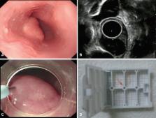
          Submucosal tumors (SMTs) originate from tissues that constitute the submucosal layer and muscularis propria, and are covered by normal mucosa. Esophageal SMTs are rare, accounting for <1% of all esophageal tumors. However, the recent widespread use of endoscopy has led to a rapid increase in incidental detection of SMTs in Korea. Esophageal SMTs are benign in ≥90% of cases, but the possibility of malignancies such as gastrointestinal stromal tumor and malignant leiomyosarcoma still exists. Therefore, patients undergo resection in the presence of symptoms or the possibility of a malignant tumor. For resection of esophageal SMTs, surgical resection was the only option available in case of possible malignancy, but minimally invasive surgery by endoscopic resection is becoming more preferable to surgical resection with the development of endoscopic ultrasonography, endoscopic techniques, and other devices.

          Related collections

          Most cited references9

          • Record: found
          • Abstract: found
          • Article: not found

          Successful outcomes of a novel endoscopic treatment for GI tumors: endoscopic submucosal dissection with a mixture of high-molecular-weight hyaluronic acid, glycerin, and sugar.

          Endoscopic submucosal dissection (ESD) has recently been developed for endoscopic treatment of GI tumors, which enables us to resect even large tumors en bloc. However, a considerable frequency of perforation has become another problem. The best way to prevent perforation is to create a sufficient submucosal fluid cushion (SFC). The aim of this study is to find out the feasibility of ESD by using a mixture of 1900 KDa hyaluronic acid (Suvenyl) and a 10% glycerin plus 5% fructose solution (Glyceol). Sixty-seven consecutive GI tumors in 54 patients who met indication criteria of ESD were enrolled. The mixing ratios of Suvenyl and Glyceol were 1:3 for esophageal/colorectal tumors and stomach tumors with scar, and 1:7 for stomach tumors without scar. After creation of SFCs, mucosal incision around the tumors and submucosal dissection under the tumors were made by cutting devices. The clinical outcomes were investigated. Mean resected and tumor sizes were 38.6 and 25.6 mm, respectively. Perforation occurred in one colon tumor with severe fibrosis (1.5%), which was managed by endoscopic clipping without salvage surgery. No blood transfusion was performed. In one stomach and in one rectal tumor (3%), endoscopic hemostasis was necessary because of postoperative bleeding. Overall endoscopic and histologic en bloc resection rates were 94% (63/67) and 78% (52/67), respectively, and there was no recurrence after follow-up of 1 year. ESD when using a mixture of Suvenyl and Glyceol results in excellent outcomes, and this injection solution should be used for ESD.
            Bookmark
            • Record: found
            • Abstract: found
            • Article: not found

            Esophageal leiomyoma: a 40-year experience.

            Esophageal leiomyomas, although infrequent, are the most common benign intramural tumors of the esophagus. They represent 10% of all gastrointestinal leiomyomas and frequently cause symptoms, necessitating resection. The Massachusetts General Hospital Pathologic Database was reviewed over a 40-year period for patients who underwent surgical resection of esophageal leiomyomas. Data analyzed included demographic information, presenting symptoms, tumor location, tumor characteristics and histology, diagnostic procedures, and treatment modalities/outcomes. Fifty-three patients were identified; 31 patients were symptomatic from their leiomyomas. Symptomatic patients presented at a mean age of 44 years old and exhibited a twofold male predominance. Mean tumor diameter among symptomatic patients was 5.3 cm, as compared to 1.5 cm in asymptomatic patients (p < 0.0001). Thirty of the symptomatic patients had solitary leiomyomas, and 1 patient had five separate leiomyomas. Eighty-four percent of the lesions in symptomatic patients occurred in the lower two-thirds of the esophagus, with epigastric discomfort being the most common presenting symptom. Among patients operated on solely for leiomyoma, 97% were enucleated without an esophageal resection. None of the leiomyomas showed malignant transformation or recurrence. All symptomatic patients had relief of symptoms, with no perioperative morbidity or mortality. In a large pathologic series, over half of all patients with esophageal leiomyomas were symptomatic. Larger tumors were significantly more likely to be symptomatic. Local enucleation by a variety of surgical approaches was accomplished in most patients. All symptomatic patients had relief of symptoms, with no perioperative morbidity or mortality. There was no observed tendency for malignant transformation or recurrence.
              Bookmark
              • Record: found
              • Abstract: not found
              • Article: not found

              Carcinoids of the rectum.

                Bookmark

                Author and article information

                Journal
                Clin Endosc
                Clin Endosc
                CE
                Clinical Endoscopy
                Korean Society of Gastrointestinal Endoscopy
                2234-2400
                2234-2443
                May 2017
                7 November 2016
                : 50
                : 3
                : 250-253
                Affiliations
                Digestive Disease Center, CHA Bundang Medical Center, CHA University, Seongnam, Korea
                Author notes
                Correspondence: Joo Young Cho Digestive Disease Center, CHA Bundang Medical Center, CHA University, 59 Yatap-ro, Bundang-gu, Seongnam 13496, Korea Tel: +82-31-780-5641, Fax: +82-31-780-5005, E-mail: cjy6695@ 123456dreamwiz.com
                Article
                ce-2016-109
                10.5946/ce.2016.109
                5475506
                27817183
                99e9459f-af12-47ed-90d1-02be09e06cb2
                Copyright © 2017 Korean Society of Gastrointestinal Endoscopy

                This is an Open Access article distributed under the terms of the Creative Commons Attribution Non-Commercial License ( http://creativecommons.org/licenses/by-nc/3.0/) which permits unrestricted non-commercial use, distribution, and reproduction in any medium, provided the original work is properly cited.

                History
                : 27 July 2016
                : 25 September 2016
                : 25 September 2016
                Categories
                Review

                Radiology & Imaging
                submucosal tumor,endosonography,endoscopic resection
                Radiology & Imaging
                submucosal tumor, endosonography, endoscopic resection

                Comments

                Comment on this article