6
views
0
recommends
+1 Recommend
0 collections
    0
    shares
      • Record: found
      • Abstract: found
      • Article: found
      Is Open Access

      A State-of-the-Art Review on the Evolving Utility of Endoscopic Ultrasound in Liver Diseases Diagnosis

      review-article

      Read this article at

      Bookmark
          There is no author summary for this article yet. Authors can add summaries to their articles on ScienceOpen to make them more accessible to a non-specialist audience.

          Abstract

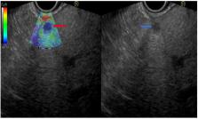
          Liver diseases are amongst the most common diseases worldwide and manifest as a parenchymatic and/or biliary injury due to several causes as well as focal liver lesions, ranging from benign to malignant ones. The diagnosis of liver diseases is based mainly on biochemical and advanced imaging studies and, when required, on liver biopsy. Endoscopic ultrasound (EUS), which combines endoscopy and ultrasonography, is one of the main examination techniques used in gastroenterology as it is applied to evaluate abnormalities in the lumen of the upper and lower gastrointestinal tract and to define pancreatic and hepato-biliary features, often in chronic patients. Given its high spatial resolution and its proximity to the liver, EUS is gaining popularity in the diagnostic work up of liver diseases. This is a comprehensive overview of the current literature on the diagnostic indications for EUS use in patients with liver diseases. We performed a MEDLINE\PubMed and Embase search, and all articles that were relevant, after reviewing abstracts, were assessed and the full text was analyzed to extract data regarding technical success, diagnostic yield, bioptic characteristics, and complications rate. EUS-guided imaging and biopsy techniques in liver diseases have shown consistent favorable promising results among the reports through the literature, with an excellent diagnostic yield and safety profile, especially in the context of focal lesions and portal hypertension. The application of EUS in the diagnosis of liver diseases is a promising technique and should be considered as a first-line therapeutic option in selected cases.

          Related collections

          Most cited references39

          • Record: found
          • Abstract: found
          • Article: not found

          Assessment of morbidity and mortality associated with EUS-guided FNA: a systematic review.

          EUS-guided FNA (EUS-FNA) permits both morphologic and cytologic analysis of lesions within or adjacent to the GI tract. Although previous studies have evaluated the accuracy of EUS-FNA, little is known about the complications of EUS-FNA. Moreover, the frequency and severity of complications may vary from center to center and may be related to differences in individual experience. To systematically review the morbidity and mortality associated with EUS-FNA. MEDLINE and EMBASE were searched to identify relevant English-language articles. EUS-FNA-specific morbidity and mortality rates. We identified 51 articles with a total of 10,941 patients who met our inclusion and exclusion criteria; the overall rate of EUS-FNA-specific morbidity was 0.98% (107/10,941). In the small proportion of patients with complications of any kind, the rates of pancreatitis (36/8246; 0.44%) and postprocedure pain (37/10,941; 0.34%) were 33.64% (36/107) and 34.58% (37/107), respectively. The mortality rate attributable to EUS-FNA-specific morbidity was 0.02% (2/10,941). Subgroup analysis showed that the morbidity rate was 2.44% in prospective studies compared with 0.35% in retrospective studies for pancreatic mass lesions (P=.000), whereas it was 2.33% versus 5.07% for pancreatic cysts (P=.036). Few articles reported well-designed, prospective studies and few focused on overall complications after EUS-FNA. EUS-FNA-related morbidity and mortality rates are relatively low, and most associated events are mild to moderate in severity. Copyright © 2011 American Society for Gastrointestinal Endoscopy. Published by Mosby, Inc. All rights reserved.
            Bookmark
            • Record: found
            • Abstract: found
            • Article: not found

            A meta-analysis of endoscopic ultrasound–fine-needle aspiration compared to endoscopic ultrasound–fine-needle biopsy: diagnostic yield and the value of onsite cytopathological assessment

            Background The diagnostic yield of endoscopic ultrasound (EUS) guided fine-needle aspiration (FNA) is variable, and partly dependent upon rapid onsite evaluation (ROSE) by a cytopathologist. Second generation fine-needle biopsy (FNB) needles are being increasingly used to obtain core histological tissue samples. Aims Studies comparing the diagnostic yield of EUS guided FNA versus FNB have reached conflicting conclusions. We therefore conducted a systematic review and meta-analysis to compare the diagnostic yield of FNA with FNB, and specifically evaluating the diagnostic value of ROSE while comparing the two types of needles. Methods We searched several databases from inception to 10 April 2016 to identify studies comparing diagnostic yield of second generation FNB needles with standard FNA needles. Risk ratios (RR) were calculated for categorical outcomes of interest (diagnostic adequacy, diagnostic accuracy, and optimal quality histological cores obtained). Standard mean difference (SMD) was calculated for continuous variables (number of passes required for diagnosis). These were pooled using random effects model of meta-analysis to account for heterogeneity. Meta-regression was conducted to evaluate the effect of ROSE on various outcomes of interest. Results Fifteen studies with a total of 1024 patients were included in the analysis. We found no significant difference in diagnostic adequacy [RR 0.98 (0.91, 1.06), (I 2 = 51 %)]. Although not statistically significant (P = 0.06), by meta-regression, in the absence of ROSE, FNB showed a relatively better diagnostic adequacy. For solid pancreatic lesions only, there was no difference in diagnostic adequacy [RR 0.96 (0.86, 1.09), (I 2 = 66 %)]. By meta-regression, in the absence of ROSE, FNB was associated with better diagnostic adequacy (P = 0.02). There was no difference in diagnostic accuracy [RR 0.99 (0.95, 1.03), (I 2 = 27 %)] or optimal quality core histological sample procurement [RR 0.97 (0.89, 1.05), (I 2 = 9.6 %)]. However, FNB established diagnosis with fewer passes [SMD 0.93 (0.45, 1.42), (I 2 = 84 %)]. The absence of ROSE was associated with a higher SMD, i. e., in the presence of an onsite pathologist, FNA required relatively fewer passes to establish the diagnosis than in the absence of an onsite pathologist. Conclusions There is no significant difference in the diagnostic yield between FNA and FNB, when FNA is accompanied by ROSE. However, in the absence of ROSE, FNB is associated with a relatively better diagnostic adequacy in solid pancreatic lesions. Also, FNB requires fewer passes to establish the diagnosis.
              Bookmark
              • Record: found
              • Abstract: found
              • Article: not found

              Transient elastography for diagnosis of advanced fibrosis and portal hypertension in patients with hepatitis C recurrence after liver transplantation.

              Recurrence of hepatitis C after liver transplantation (LT) is the main cause of graft loss and retransplantation. Frequent liver biopsies are essential to follow-up hepatitis C virus (HCV)-induced liver damage. However, liver biopsy is an invasive and expensive procedure. We evaluated prospectively the diagnostic accuracy of noninvasive measurement of liver stiffness (by transient elastography) to assess the severity of hepatitis C recurrence after LT. For this purpose, we included 124 HCV-infected liver transplant recipients who underwent 169 liver biopsies and 129 hepatic hemodynamic studies with determination of hepatic venous pressure gradient (HVPG). Simultaneously, patients underwent measurement of liver stiffness. Liver fibrosis was mild (F0-F1) in 96 cases (57%) and significant (F2-F4) in 73 (43%). HVPG was normal ( or=6 mm Hg) in 60 (46%). Using a liver stiffness cutoff value of 8.5 kilopascals, the sensitivity, specificity, negative predictive value, and positive predictive value for diagnosis of fibrosis >or=F2 were 90%, 81%, 79%, and 92%, respectively. The area under the curve (AUC) for diagnosis of fibrosis >or=F2, >or=F3 and F4 were 0.90, 0.93, and 0.98, respectively. There was a close direct correlation between liver stiffness and HVPG (Pearson coefficient, 0.84; P or=6 mm Hg) was 0.93. Importantly, none of the individuals with liver stiffness below the cutoff value had either bridging fibrosis (F3) or cirrhosis (F4) or significant portal hypertension (HVPG >or=10 mm Hg). In conclusion, determination of liver stiffness is an extremely valuable tool to assess the severity of HCV recurrence after LT and in reducing the need of follow-up liver biopsies.
                Bookmark

                Author and article information

                Journal
                Diagnostics (Basel)
                Diagnostics (Basel)
                diagnostics
                Diagnostics
                MDPI
                2075-4418
                23 July 2020
                August 2020
                : 10
                : 8
                : 512
                Affiliations
                [1 ]Department of Gastroenterology, Galilee Medical Center, Nahariya 22100, Israel; wisams@ 123456gmc.gov.il (W.S.); anas18kadah@ 123456msn.com (A.K.)
                [2 ]Faculty of Medicine in the Galilee, Bar-Ilan University, Safed 1311502, Israel
                [3 ]Department of Gastroenterology, Sharee Zedek Medical Center, Jerusalem 9103102, Israel; mahmudmahamid@ 123456yahoo.com
                [4 ]Gastroenterology Unit, Molinette Hospital, 10126 Turin, Italy; rinaldo_pellican@ 123456hotmail.com
                [5 ]Gastroenterology and Endoscopy Units, The Nazareth Hospital, EMMS, Nazareth 16100, Israel; amir.mari@ 123456hotmail.com
                Author notes
                [* ]Correspondence: tawfikkhoury1@ 123456hotmail.com ; Tel.: +972-509870611
                Author information
                https://orcid.org/0000-0003-3438-0649
                https://orcid.org/0000-0001-6699-8625
                Article
                diagnostics-10-00512
                10.3390/diagnostics10080512
                7459648
                32717886
                991d0cc2-0e88-445e-86a8-6f0c193ae084
                © 2020 by the authors.

                Licensee MDPI, Basel, Switzerland. This article is an open access article distributed under the terms and conditions of the Creative Commons Attribution (CC BY) license ( http://creativecommons.org/licenses/by/4.0/).

                History
                : 06 July 2020
                : 21 July 2020
                Categories
                Review

                eus,endoscopic ultrasonography,liver diseases,diagnosis
                eus, endoscopic ultrasonography, liver diseases, diagnosis

                Comments

                Comment on this article