54
views
0
recommends
+1 Recommend
0 collections
    0
    shares
      • Record: found
      • Abstract: found
      • Article: found
      Is Open Access

      Early ERK1/2 activation promotes DRP1-dependent mitochondrial fission necessary for cell reprogramming

      research-article

      Read this article at

      Bookmark
          There is no author summary for this article yet. Authors can add summaries to their articles on ScienceOpen to make them more accessible to a non-specialist audience.

          Abstract

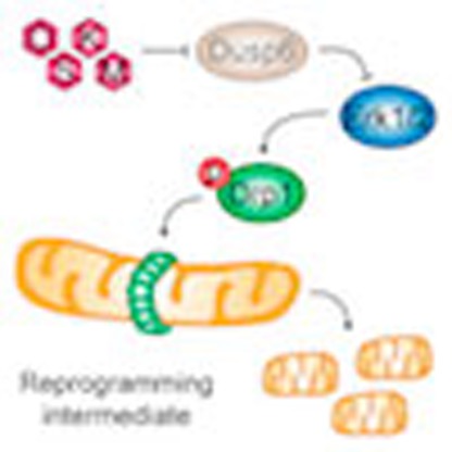
          During the process of reprogramming to induced pluripotent stem (iPS) cells, somatic cells switch from oxidative to glycolytic metabolism, a transition associated with profound mitochondrial reorganization. Neither the importance of mitochondrial remodelling for cell reprogramming, nor the molecular mechanisms controlling this process are well understood. Here, we show that an early wave of mitochondrial fragmentation occurs upon expression of reprogramming factors. Reprogramming-induced mitochondrial fission is associated with a minor decrease in mitochondrial mass but not with mitophagy. The pro-fission factor Drp1 is phosphorylated early in reprogramming, and its knockdown and inhibition impairs both mitochondrial fragmentation and generation of iPS cell colonies. Drp1 phosphorylation depends on Erk activation in early reprogramming, which occurs, at least in part, due to downregulation of the MAP kinase phosphatase Dusp6. Taken together, our data indicate that mitochondrial fission controlled by an Erk-Drp1 axis constitutes an early and necessary step in the reprogramming process to pluripotency.

          Abstract

          Reprogramming of somatic cells is a stepwise process where cells must overcome several barriers before reaching the pluripotent state. Here the authors show that mitochondrial fission in response to ERK1/2 signalling is an important early step during reprogramming to pluripotency.

          Related collections

          Most cited references34

          • Record: found
          • Abstract: found
          • Article: not found

          Roles of the mammalian mitochondrial fission and fusion mediators Fis1, Drp1, and Opa1 in apoptosis.

          During apoptosis, the mitochondrial network fragments. Using short hairpin RNAs for RNA interference, we manipulated the expression levels of the proteins hFis1, Drp1, and Opa1 that are involved in mitochondrial fission and fusion in mammalian cells, and we characterized their functions in mitochondrial morphology and apoptosis. Down-regulation of hFis1 powerfully inhibits cell death to an extent significantly greater than down-regulation of Drp1 and at a stage of apoptosis distinct from that induced by Drp1 inhibition. Cells depleted of Opa1 are extremely sensitive to exogenous apoptosis induction, and some die spontaneously by a process that requires hFis1 expression. Wild-type Opa1 may function normally as an antiapoptotic protein, keeping spontaneous apoptosis in check. However, if hFis1 is down-regulated, cells do not require Opa1 to prevent apoptosis, suggesting that Opa1 may be normally counteracting the proapoptotic action of hFis1. We also demonstrate in this study that mitochondrial fragmentation per se does not result in apoptosis. However, we provide further evidence that multiple components of the mitochondrial morphogenesis machinery can positively and negatively regulate apoptosis.
            Bookmark
            • Record: found
            • Abstract: found
            • Article: not found

            FGF stimulation of the Erk1/2 signalling cascade triggers transition of pluripotent embryonic stem cells from self-renewal to lineage commitment.

            Pluripotent embryonic stem (ES) cells must select between alternative fates of self-replication and lineage commitment during continuous proliferation. Here, we delineate the role of autocrine production of fibroblast growth factor 4 (Fgf4) and associated activation of the Erk1/2 (Mapk3/1) signalling cascade. Fgf4 is the major stimulus activating Erk in mouse ES cells. Interference with FGF or Erk activity using chemical inhibitors or genetic ablations does not impede propagation of undifferentiated ES cells. Instead, such manipulations restrict the ability of ES cells to commit to differentiation. ES cells lacking Fgf4 or treated with FGF receptor inhibitors resist neural and mesodermal induction, and are refractory to BMP-induced non-neural differentiation. Lineage commitment potential of Fgf4-null cells is restored by provision of FGF protein. Thus, FGF enables rather than antagonises the differentiation activity of BMP. The key downstream role of Erk signalling is revealed by examination of Erk2-null ES cells, which fail to undergo either neural or mesodermal differentiation in adherent culture, and retain expression of pluripotency markers Oct4, Nanog and Rex1. These findings establish that Fgf4 stimulation of Erk1/2 is an autoinductive stimulus for naïve ES cells to exit the self-renewal programme. We propose that the Erk cascade directs transition to a state that is responsive to inductive cues for germ layer segregation. Consideration of Erk signalling as a primary trigger that potentiates lineage commitment provides a context for reconciling disparate views on the contribution of FGF and BMP pathways during germ layer specification in vertebrate embryos.
              Bookmark
              • Record: found
              • Abstract: found
              • Article: not found

              The mitochondrial protein hFis1 regulates mitochondrial fission in mammalian cells through an interaction with the dynamin-like protein DLP1.

              The yeast protein Fis1p has been shown to participate in mitochondrial fission mediated by the dynamin-related protein Dnm1p. In mammalian cells, the dynamin-like protein DLP1/Drp1 functions as a mitochondrial fission protein, but the mechanisms by which DLP1/Drp1 and the mitochondrial membrane interact during the fission process are undefined. In this study, we have tested the role of a mammalian homologue of Fis1p, hFis1, and provided new and mechanistic information about the control of mitochondrial fission in mammalian cells. Through differential tagging and deletion experiments, we demonstrate that the intact C-terminal structure of hFis1 is essential for mitochondrial localization, whereas the N-terminal region of hFis1 is necessary for mitochondrial fission. Remarkably, an increased level of cellular hFis1 strongly promotes mitochondrial fission, resulting in an accumulation of fragmented mitochondria. Conversely, cell microinjection of hFis1 antibodies or treatment with hFis1 antisense oligonucleotides induces an elongated and collapsed mitochondrial morphology. Further, fluorescence resonance energy transfer and coimmunoprecipitation studies demonstrate that hFis1 interacts with DLP1. These results suggest that hFis1 participates in mitochondrial fission through an interaction that recruits DLP1 from the cytosol. We propose that hFis1 is a limiting factor in mitochondrial fission and that the number of hFis1 molecules on the mitochondrial surface determines fission frequency.
                Bookmark

                Author and article information

                Journal
                Nat Commun
                Nat Commun
                Nature Communications
                Nature Publishing Group
                2041-1723
                31 March 2016
                2016
                : 7
                : 11124
                Affiliations
                [1 ]Departamento de Biología Celular, Universidad de Valencia , Burjassot 46100, Spain
                [2 ]Departamento de Bioquímica y Biología Molecular, Universidad de Valencia , Burjassot 46100, Spain
                [3 ]Unidad de Hepatología Experimental, CIBERehd, Instituto de Investigación Sanitaria La Fe , Valencia 46026, Spain
                [4 ]Centre de Medicina Regenerativa de Barcelona , Barcelona 08003, Spain
                [5 ]Centro de Investigación Biomédica en Red en Bioingeniería, Biomateriales y Nanomedicina , Madrid 28029, Spain
                [6 ]Institució Catalana de Recerca i Estudis Avançats , Barcelona 08010, Spain
                Author notes
                Author information
                http://orcid.org/0000-0003-2189-9775
                http://orcid.org/0000-0001-6906-7164
                Article
                ncomms11124
                10.1038/ncomms11124
                4821885
                27030341
                96e0dc9b-e82e-4d24-a54a-bcff41efa898
                Copyright © 2016, Nature Publishing Group, a division of Macmillan Publishers Limited. All Rights Reserved.

                This work is licensed under a Creative Commons Attribution 4.0 International License. The images or other third party material in this article are included in the article's Creative Commons license, unless indicated otherwise in the credit line; if the material is not included under the Creative Commons license, users will need to obtain permission from the license holder to reproduce the material. To view a copy of this license, visit http://creativecommons.org/licenses/by/4.0/

                History
                : 07 July 2015
                : 23 February 2016
                Categories
                Article

                Uncategorized
                Uncategorized

                Comments

                Comment on this article