21
views
0
recommends
+1 Recommend
0 collections
    0
    shares
      • Record: found
      • Abstract: found
      • Article: found
      Is Open Access

      Development of an Equine Groove Model to Induce Metacarpophalangeal Osteoarthritis: A Pilot Study on 6 Horses

      research-article

      Read this article at

      Bookmark
          There is no author summary for this article yet. Authors can add summaries to their articles on ScienceOpen to make them more accessible to a non-specialist audience.

          Abstract

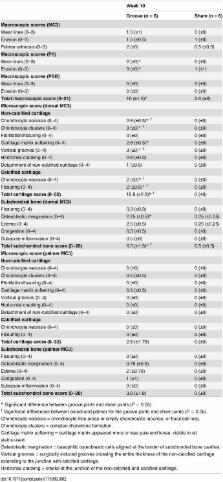
          The aim of this work was to develop an equine metacarpophalangeal joint model that induces osteoarthritis that is not primarily mediated by instability or inflammation. The study involved six Standardbred horses. Standardized cartilage surface damage or “grooves” were created arthroscopically on the distal dorsal aspect of the lateral and medial metacarpal condyles of a randomly chosen limb. The contralateral limb was sham operated. After 2 weeks of stall rest, horses were trotted 30 minutes every other day for 8 weeks, then evaluated for lameness and radiographed. Synovial fluid was analyzed for cytology and biomarkers. At 10 weeks post-surgery, horses were euthanized for macroscopic and histologic joint evaluation. Arthroscopic grooving allowed precise and identical damage to the cartilage of all animals. Under the controlled exercise regime, this osteoarthritis groove model displayed significant radiographic, macroscopic, and microscopic degenerative and reactive changes. Histology demonstrated consistent surgically induced grooves limited to non-calcified cartilage and accompanied by secondary adjacent cartilage lesions, chondrocyte necrosis, chondrocyte clusters, cartilage matrix softening, fissuring, mild subchondral bone inflammation, edema, and osteoblastic margination. Synovial fluid biochemistry and cytology demonstrated significantly elevated total protein without an increase in prostaglandin E2, neutrophils, or chondrocytes. This equine metacarpophalangeal groove model demonstrated that standardized non-calcified cartilage damage accompanied by exercise triggered altered osteochondral morphology and cartilage degeneration with minimal or inefficient repair and little inflammatory response. This model, if validated, would allow for assessment of disease processes and the effects of therapy.

          Related collections

          Most cited references26

          • Record: found
          • Abstract: found
          • Article: not found

          Treatment of experimental equine osteoarthritis by in vivo delivery of the equine interleukin-1 receptor antagonist gene.

          Osteoarthritis in horses and in humans is a significant social and economic problem and continued research and improvements in therapy are needed. Because horses have naturally occurring osteoarthritis, which is similar to that of humans, the horse was chosen as a species with which to investigate gene transfer as a potential therapeutic modality for the clinical treatment of osteoarthritis. Using an established model of equine osteoarthritis that mimics clinical osteoarthritis, the therapeutic effects resulting from intra-articular overexpression of the equine interleukin-1 receptor antagonist gene through adenoviral-mediated gene transfer were investigated. In vivo delivery of the equine IL-IRa gene led to elevated intra-articular expression of interleukin-1 receptor antagonist for approximately 28 days, resulting in significant improvement in clinical parameters of pain and disease activity, preservation of articular cartilage, and beneficial effects on the histologic parameters of synovial membrane and articular cartilage. Based on these findings, gene transfer of interleukin-1 receptor antagonist is an attractive treatment modality for the equine patient and also offers future promise for human patients with osteoarthritis.
            Bookmark
            • Record: found
            • Abstract: found
            • Article: not found

            Early events in cartilage repair after subchondral bone microfracture.

            The current study investigated healing of large full-thickness articular cartilage defects during the first 8 weeks with and without penetration of the subchondral bone using microfracture in an established equine model of cartilage healing. Chondral defects in the weightbearing portion of the medial femoral condyle were made bilaterally; one defect in each horse was microfractured whereas the contralateral leg served as the control. The expression of cartilage extracellular matrix components (Types I and II collagen and aggrecan) was evaluated using histologic techniques, reverse transcription coupled polymerase chain reaction, in situ hybridization, and immunohistochemistry. This study confirms an increase in Type II collagen mRNA expression in repair tissue as early as 6 weeks after microfracture. Although other matrix mRNA and protein levels changed in concentration and tissue location over the course of the study, no significant differences were seen in microfractured defects. Although the microfracture techniques appear to improve clinical functionality, volume of repair tissue, and augment Type II collagen content, aggrecan content is less than ideal. Therefore, methods to enhance key matrix components such as aggrecan after microfracture may additionally improve repair tissue observed after the procedure.
              Bookmark
              • Record: found
              • Abstract: found
              • Article: not found

              Changes in synovial fluid and serum biomarkers with exercise and early osteoarthritis in horses.

              To discriminate between changes in biomarkers with exercise compared to changes in biomarkers with osteoarthritis (OA) in exercising horses. Sixteen, 2-year-old horses were randomly assigned either to an exercise-alone (n=8) or OA-affected (also exercised) (n=8) group. All horses had both mid-carpal joints arthroscoped and OA induced in one mid-carpal joint in the OA-affected joints of OA-affected horses. Two weeks after surgery all horses commenced a strenuous exercise program on a high-speed treadmill. Clinical outcomes and synovial fluid and serum biomarkers, were evaluated weekly. Synovial and serum biomarkers evaluated were epitope CS846 (CS846), epitope CPII (CPII), glycosaminoglycans (GAGs), epitope Col CEQ (Col CEQ) (a marker of type II collagen degradation), type I and II collagen degradation fragments (C1,2C), osteocalcin, C-terminal of bone type I collagen (CTX1), type I collagen (Col I) and (synovial fluid only of cartilage) prostaglandin E2 (PGE2) levels. Horses were euthanized at day 91 and their joints assessed grossly, histopathologically, and histochemically. Exercise induced a significant increase in synovial fluid CS846, CPII, GAG, Col CEQ, C1,2C, osteocalcin and Col I concentrations. There was a significant increase in synovial fluid CS846, CPII, Col CEQ, C1,2C, osteocalcin, Col I and PGE2 concentrations in OA-affected joints compared to exercise-alone joints. The concentration of serum CS846, CPII, GAG, osteocalcin, C1,2C and Col I increased with exercise. For each of these biomarkers there was also a statistically significant increase in serum biomarker levels in OA-affected horses compared to exercise-alone horses. Six synovial fluid and serum biomarkers were useful in separating early experimental OA from exercise alone but synovial fluid CTX1 and serum Col CEQ and CTX1 were not.
                Bookmark

                Author and article information

                Contributors
                Role: Academic Editor
                Journal
                PLoS One
                PLoS ONE
                plos
                plosone
                PLoS ONE
                Public Library of Science (San Francisco, CA USA )
                1932-6203
                2015
                13 February 2015
                : 10
                : 2
                : e0115089
                Affiliations
                [1 ]Equine Research Centre, University of Lyon, VetAgro Sup, Veterinary Campus of Lyon, GREMERES-ICE, Marcy l’Etoile, France
                [2 ]Exploratory Unit, Sanofi-aventis Recherche, Montpellier, France
                [3 ]Clinical unit for osteoarticular diseases, CHU Lapeyronie University Hospital, Montpellier, France
                Instituto Butantan, BRAZIL
                Author notes

                Competing Interests: SH, BR, FM, GP, PC, RS are employed by a commercial company Sanofi-aventis Recherche. This does not alter the authors’ adherence to all the PLOS ONE policies on sharing data and materials.

                Conceived and designed the experiments: UM OML MG GP RS. Performed the experiments: UM OML MG. Analyzed the data: UM SH BR FM GP ES PC RS. Contributed reagents/materials/analysis tools: UM OML MG SH BR FM GP PC RS. Wrote the paper: UM OML MG SH BR FM GP ES PC CJ RS.

                ‡ UM, OML, MG, and RS contributed equally to this work. SH, BR, FM, and GP also contributed equally to this work.

                Article
                PONE-D-14-39881
                10.1371/journal.pone.0115089
                4332493
                25680102
                93682a30-3fe6-46ae-a0c0-34b454e4b9ae
                Copyright @ 2015

                This is an open access article distributed under the terms of the Creative Commons Attribution License, which permits unrestricted use, distribution, and reproduction in any medium, provided the original author and source are credited

                History
                : 4 September 2014
                : 10 November 2014
                Page count
                Figures: 5, Tables: 2, Pages: 18
                Funding
                The research was supported by GREMERES (Groupe de Recherche en Médecine et Rééducation des Equidés de Sport), Veterinary Campus of Lyon (VetAgro Sup) and the Exploratory unit at Sanofi-Aventis contract service. SH, BR, FM, GP, PC, RS received funding from Sanofi-Aventis in the form of salary. The funders had no role in study design, data collection and analysis, decision to publish, or preparation of the manuscript.
                Categories
                Research Article
                Custom metadata
                All relevant data are within the paper.

                Uncategorized
                Uncategorized

                Comments

                Comment on this article