56
views
0
recommends
+1 Recommend
0 collections
    0
    shares
      • Record: found
      • Abstract: found
      • Article: found
      Is Open Access

      Centronuclear (myotubular) myopathy

      review-article

      Read this article at

      Bookmark
          There is no author summary for this article yet. Authors can add summaries to their articles on ScienceOpen to make them more accessible to a non-specialist audience.

          Abstract

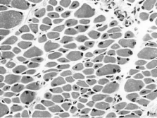
          Centronuclear myopathy (CNM) is an inherited neuromuscular disorder characterised by clinical features of a congenital myopathy and centrally placed nuclei on muscle biopsy.

          The incidence of X-linked myotubular myopathy is estimated at 2/100000 male births but epidemiological data for other forms are not currently available.

          The clinical picture is highly variable. The X-linked form usually gives rise to a severe phenotype in males presenting at birth with marked weakness and hypotonia, external ophthalmoplegia and respiratory failure. Signs of antenatal onset comprise reduced foetal movements, polyhydramnios and thinning of the ribs on chest radiographs; birth asphyxia may be the present. Affected infants are often macrosomic, with length above the 90 th centile and large head circumference. Testes are frequently undescended. Both autosomal-recessive (AR) and autosomal-dominant (AD) forms differ from the X-linked form regarding age at onset, severity, clinical characteristics and prognosis. In general, AD forms have a later onset and milder course than the X-linked form, and the AR form is intermediate in both respects.

          Mutations in the myotubularin (MTM1) gene on chromosome Xq28 have been identified in the majority of patients with the X-linked recessive form, whilst AD and AR forms have been associated with mutations in the dynamin 2 (DNM2) gene on chromosome 19p13.2 and the amphiphysin 2 (BIN1) gene on chromosome 2q14, respectively. Single cases with features of CNM have been associated with mutations in the skeletal muscle ryanodine receptor (RYR1) and the hJUMPY (MTMR14) genes.

          Diagnosis is based on typical histopathological findings on muscle biopsy in combination with suggestive clinical features; muscle magnetic resonance imaging may complement clinical assessment and inform genetic testing in cases with equivocal features. Genetic counselling should be offered to all patients and families in whom a diagnosis of CNM has been made.

          The main differential diagnoses include congenital myotonic dystrophy and other conditions with severe neonatal hypotonia.

          Management of CNM is mainly supportive, based on a multidisciplinary approach. Whereas the X-linked form due to MTM1 mutations is often fatal in infancy, dominant forms due to DNM2 mutations and some cases of the recessive BIN1-related form appear to be associated with an overall more favourable prognosis.

          Related collections

          Most cited references144

          • Record: found
          • Abstract: found
          • Article: not found

          BAR domains as sensors of membrane curvature: the amphiphysin BAR structure.

          The BAR (Bin/amphiphysin/Rvs) domain is the most conserved feature in amphiphysins from yeast to human and is also found in endophilins and nadrins. We solved the structure of the Drosophila amphiphysin BAR domain. It is a crescent-shaped dimer that binds preferentially to highly curved negatively charged membranes. With its N-terminal amphipathic helix and BAR domain (N-BAR), amphiphysin can drive membrane curvature in vitro and in vivo. The structure is similar to that of arfaptin2, which we find also binds and tubulates membranes. From this, we predict that BAR domains are in many protein families, including sorting nexins, centaurins, and oligophrenins. The universal and minimal BAR domain is a dimerization, membrane-binding, and curvature-sensing module.
            Bookmark
            • Record: found
            • Abstract: found
            • Article: not found

            Amphiphysin 2 (Bin1) and T-tubule biogenesis in muscle.

            In striated muscle, the plasma membrane forms tubular invaginations (transverse tubules or T-tubules) that function in depolarization-contraction coupling. Caveolin-3 and amphiphysin were implicated in their biogenesis. Amphiphysin isoforms have a putative role in membrane deformation at endocytic sites. An isoform of amphiphysin 2 concentrated at T-tubules induced tubular plasma membrane invaginations when expressed in nonmuscle cells. This property required exon 10, a phosphoinositide-binding module. In developing myotubes, amphiphysin 2 and caveolin-3 segregated in tubular and vesicular portions of the T-tubule system, respectively. These findings support a role of the bilayer-deforming properties of amphiphysin at T-tubules and, more generally, a physiological role of amphiphysin in membrane deformation.
              Bookmark
              • Record: found
              • Abstract: found
              • Article: not found

              A gene mutated in X-linked myotubular myopathy defines a new putative tyrosine phosphatase family conserved in yeast.

              X-linked recessive myotubular myopathy (MTM1) is characterized by severe hypotonia and generalized muscle weakness, with impaired maturation of muscle fibres. We have restricted the candidate region to 280 kb and characterized two candidate genes using positional cloning strategies. The presence of frameshift or missense mutations (of which two are new mutations) in seven patients proved that one of these genes is indeed implicated in MTM1. The protein encoded by the MTM1 gene is highly conserved in yeast, which is surprising for a muscle specific disease. The protein contains the consensus sequence for the active site of tyrosine phosphatases, a wide class of proteins involved in signal transduction. At least three other genes, one located within 100 kb distal from the MTM1 gene, encode proteins with very high sequence similarities and define, together with the MTM1 gene, a new family of putative tyrosine phosphatases in man.
                Bookmark

                Author and article information

                Journal
                Orphanet J Rare Dis
                Orphanet Journal of Rare Diseases
                BioMed Central
                1750-1172
                2008
                25 September 2008
                : 3
                : 26
                Affiliations
                [1 ]Department of Paediatric Neurology, Neuromuscular Service, Evelina Children's Hospital, St Thomas' Hospital, Lambeth Palace Road, London, SE1 7EH, UK
                [2 ]Clinical Neuroscience Division, King's College, London, UK
                [3 ]Department of Medical Genetics, University of Helsinki, Helsinki, Finland
                [4 ]The Folkhälsan Department of Medical Genetics, Helsinki, Finland
                [5 ]Department of Neurobiology and Genetics, IGBMC (Institut de Génétique et de Biologie Moléculaire et Cellulaire), Illkirch F-67400, France
                [6 ]Inserm, U596, Illkirch F-67400, France
                [7 ]CNRS, UMR7104, Illkirch F-67400, France
                [8 ]Université Louis Pasteur, Strasbourg F-67000, France
                [9 ]Collège de France, Illkirch F-67400, France
                Article
                1750-1172-3-26
                10.1186/1750-1172-3-26
                2572588
                18817572
                92497581-0270-4cba-9395-5ec5006f64bf
                Copyright © 2008 Jungbluth et al; licensee BioMed Central Ltd.

                This is an Open Access article distributed under the terms of the Creative Commons Attribution License ( http://creativecommons.org/licenses/by/2.0), which permits unrestricted use, distribution, and reproduction in any medium, provided the original work is properly cited.

                History
                : 22 April 2008
                : 25 September 2008
                Categories
                Review

                Infectious disease & Microbiology
                Infectious disease & Microbiology

                Comments

                Comment on this article