6
views
0
recommends
+1 Recommend
0 collections
    0
    shares
      • Record: found
      • Abstract: found
      • Article: found
      Is Open Access

      Bio-inspired, bio-degradable adenosine 5′-diphosphate-modified hyaluronic acid coordinated hydrophobic undecanal-modified chitosan for hemostasis and wound healing

      research-article

      Read this article at

      Bookmark
          There is no author summary for this article yet. Authors can add summaries to their articles on ScienceOpen to make them more accessible to a non-specialist audience.

          Abstract

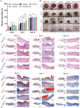
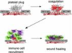
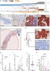
          Uncontrolled hemorrhage and wound infection are crucial causes of trauma-associated death in both the military and the clinic. Therefore, developing an efficient and rapid hemostatic method with biocompatibility, easy degradation, and wound healing is of great importance and desirability. Inspired by spontaneous blood cell plug formation in the hemostasis process, an adenosine 5′-diphosphate modified pro-coagulation hyaluronic acid (HA-ADP) coordinated with enhanced antibacterial activity of undecanal-modified chitosan (UCS) was fabricated through physical electrostatic cross-linking and freeze-drying. The as-prepared hydrogel sponges showed a porous structure suitable for blood cell adhesion. In particular, the hydrogel exhibited excellent antibacterial ability and promoted the adhesion of platelets and red blood cells, thus inducing a prominent pro-coagulation ability via platelet activation, which exhibits a shorter hemostasis time (58.94% of control) in vitro. Compared with commercially available CELOX and gelatin sponge (GS), HA-ADP/UCS accelerates hemostasis and reduces blood loss in both rat tail amputation and rat artery injury models. Furthermore, all the samples exhibited superior cytocompatibility and biodegradability. Due to these performances, HA-ADP/UCS promoted full-thickness skin defect healing significantly in vivo. All the properties of HA-ADP/UCS suggest that it has great potential for translation as a clinical application material for hemostatic and wound healing.

          Graphical abstract

          Highlights

          • Using adenosine 5'-diphosphate, a physiologically platelet agonist, to modify hyaluronic acids to promote hemostatic effect.

          • Using undecanal to modify Chitosan fabricated with HA-ADP via electrostatic interactions and noncovalent crosslinking method.

          • The hydrogel sponges have excellent antibacterial properties related to the bacterial disruption abilities of UCS.

          • HA-ADP/UCS posed a great hemostatic performance, promoting wound healing by regulating inflammatory response in early stage.

          Related collections

          Most cited references49

          • Record: found
          • Abstract: found
          • Article: found
          Is Open Access

          Platelet secretion: From haemostasis to wound healing and beyond

          Upon activation, platelets secrete more than 300 active substances from their intracellular granules. Platelet dense granule components, such as ADP and polyphosphates, contribute to haemostasis and coagulation, but also play a role in cancer metastasis. α-Granules contain multiple cytokines, mitogens, pro- and anti-inflammatory factors and other bioactive molecules that are essential regulators in the complex microenvironment of the growing thrombus but also contribute to a number of disease processes. Our understanding of the molecular mechanisms of secretion and the genetic regulation of granule biogenesis still remains incomplete. In this review we summarise our current understanding of the roles of platelet secretion in health and disease, and discuss some of the hypotheses that may explain how platelets may control the release of its many secreted components in a context-specific manner, to allow platelets to play multiple roles in health and disease.
            Bookmark
            • Record: found
            • Abstract: not found
            • Article: not found

            Physical Double‐Network Hydrogel Adhesives with Rapid Shape Adaptability, Fast Self‐Healing, Antioxidant and NIR/pH Stimulus‐Responsiveness for Multidrug‐Resistant Bacterial Infection and Removable Wound Dressing

              Bookmark
              • Record: found
              • Abstract: found
              • Article: found
              Is Open Access

              Prostate-specific Membrane Antigen Heterogeneity and DNA Repair Defects in Prostate Cancer

              Background Prostate-specific membrane antigen (PSMA; folate hydrolase) prostate cancer (PC) expression has theranostic utility. Objective To elucidate PC PSMA expression and associate this with defective DNA damage repair (DDR). Design, setting, and participants Membranous PSMA (mPSMA) expression was scored immunohistochemically from metastatic castration-resistant PC (mCRPC) and matching, same-patient, diagnostic biopsies, and correlated with next-generation sequencing (NGS) and clinical outcome data. Outcome measurements and statistical analysis Expression of mPSMA was quantitated by modified H-score. Patient DNA was tested by NGS. Gene expression and activity scores were determined from mCRPC transcriptomes. Statistical correlations utilised Wilcoxon signed rank tests, survival was estimated by Kaplan-Meier test, and sample heterogeneity was quantified by Shannon's diversity index. Results and limitations Expression of mPSMA at diagnosis was associated with higher Gleason grade (p = 0.04) and worse overall survival (p = 0.006). Overall, mPSMA expression levels increased at mCRPC (median H-score [interquartile range]: castration-sensitive prostate cancer [CSPC] 17.5 [0.0–60.0] vs mCRPC 55.0 [2.8–117.5]). Surprisingly, 42% (n = 16) of CSPC and 27% (n = 16) of mCRPC tissues sampled had no detectable mPSMA (H-score <10). Marked intratumour heterogeneity of mPSMA expression, with foci containing no detectable PSMA, was observed in all mPSMA expressing CSPC (100%) and 37 (84%) mCRPC biopsies. Heterogeneous intrapatient mPSMA expression between metastases was also observed, with the lowest expression in liver metastases. Tumours with DDR had higher mPSMA expression (p = 0.016; 87.5 [25.0–247.5] vs 20 [0.3–98.8]; difference in medians 60 [5.0–95.0]); validation cohort studies confirmed higher mPSMA expression in patients with deleterious aberrations in BRCA2 (p < 0.001; median H-score: 300 [165–300]; difference in medians 195.0 [100.0–270.0]) and ATM (p = 0.005; 212.5 [136.3–300]; difference in medians 140.0 [55.0–200]) than in molecularly unselected mCRPC biopsies (55.0 [2.75–117.5]). Validation studies using mCRPC transcriptomes corroborated these findings, also indicating that SOX2 high tumours have low PSMA expression. Conclusions Membranous PSMA expression is upregulated in some but not all PCs, with mPSMA expression demonstrating marked inter- and intrapatient heterogeneity. DDR aberrations are associated with higher mPSMA expression and merit further evaluation as predictive biomarkers of response for PSMA-targeted therapies in larger, prospective cohorts. Patient summary Through analysis of prostate cancer samples, we report that the presence of prostate-specific membrane antigen (PSMA) is extremely variable both within one patient and between different patients. This may limit the usefulness of PSMA scans and PSMA-targeted therapies. We show for the first time that prostate cancers with defective DNA repair produce more PSMA and so may respond better to PSMA-targeting treatments.
                Bookmark

                Author and article information

                Contributors
                Journal
                Bioact Mater
                Bioact Mater
                Bioactive Materials
                KeAi Publishing
                2452-199X
                29 January 2022
                November 2022
                29 January 2022
                : 17
                : 162-177
                Affiliations
                [a ]Shanghai Key Laboratory of Orthopedic Implant, Department of Orthopedic Surgery, Shanghai Ninth People's Hospital, Shanghai Jiao Tong University School of Medicine, No. 639 Zhizaoju Rd, Shanghai, 200011, China
                [b ]Department of Orthopaedic Surgery, Children's Hospital of Nanjing Medical University, No. 72 Guangzhou Rd, Nanjing, 210008, China
                [c ]Department of Rehabilitation, Huashan Hospital, Fudan University, No.12 Middle Urumqi Rd, Shanghai, 200040, China
                [d ]Med-X Research Institute, School of Biomedical Engineering, Shanghai Jiao Tong University, No. 1956 Huashan Rd, Shanghai, 200030, China
                [e ]Department of Orthopedics, Shanghai General Hospital, Shanghai Jiao Tong University School of Medicine, No. 85 Wujin Rd, Shanghai, 200080, China
                Author notes
                []Corresponding authorShanghai Key Laboratory of Orthopedic Implant, Department of Orthopedic Surgery, Shanghai Ninth People's Hospital, Shanghai Jiao Tong University School of Medicine, No. 639 Zhizaoju Rd, Shanghai, 200011, China. wangjw-team@ 123456shsmu.edu.cn
                [∗∗ ]Corresponding author.
                [∗∗∗ ]Corresponding author.
                [∗∗∗∗ ]Corresponding author.
                [1]

                Y.L. and H.N. and C.W. contributed equally to the work.

                Article
                S2452-199X(22)00033-0
                10.1016/j.bioactmat.2022.01.025
                8965034
                35386451
                8ec2e433-48e9-4532-afff-e9ba19fe0b3e
                © 2022 The Authors

                This is an open access article under the CC BY-NC-ND license (http://creativecommons.org/licenses/by-nc-nd/4.0/).

                History
                : 4 November 2021
                : 13 January 2022
                : 13 January 2022
                Categories
                Article

                hemostasis,antibacterial,wound healing,hyaluronic acid,chitosan

                Comments

                Comment on this article