43
views
0
recommends
+1 Recommend
0 collections
    0
    shares
      • Record: found
      • Abstract: found
      • Article: not found

      RBM3 mediates structural plasticity and protective effects of cooling in neurodegeneration

      research-article

      Read this article at

      ScienceOpenPublisherPMC
      Bookmark
          There is no author summary for this article yet. Authors can add summaries to their articles on ScienceOpen to make them more accessible to a non-specialist audience.

          Abstract

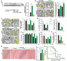
          In the healthy adult brain synapses are continuously remodelled through a process of elimination and formation known as structural plasticity 1 . Reduction in synapse number is a consistent early feature of neurodegenerative diseases 2, 3 , suggesting deficient compensatory mechanisms. While much is known about toxic processes leading to synaptic dysfunction and loss in these disorders 2, 3 , how synaptic regeneration is affected is unknown. In hibernating mammals, cooling induces loss of synaptic contacts, which are reformed on rewarming, a form of structural plasticity 4, 5 . We have found that similar changes occur in artificially cooled laboratory rodents. Cooling and hibernation also induce a number cold-shock proteins in the brain, including the RNA binding protein, RBM3 6 . The relationship of such proteins to structural plasticity is unknown. Here we show that synapse regeneration is impaired in mouse models of neurodegenerative disease, in association with the failure to induce RBM3. In both prion-infected and 5×FAD (Alzheimer-type) mice 7 , the capacity to regenerate synapses after cooling declined in parallel with the loss of induction of RBM3. Enhanced expression of RBM3 in the hippocampus prevented this deficit and restored the capacity for synapse reassembly after cooling. Further, RBM3 over-expression, achieved either by boosting endogenous levels through hypothermia prior to the loss of the RBM3 response, or by lentiviral delivery, resulted in sustained synaptic protection in 5×FAD mice and throughout the course of prion disease, preventing behavioural deficits and neuronal loss and significantly prolonging survival. In contrast, knockdown of RBM3 exacerbated synapse loss in both models and accelerated disease and prevented the neuroprotective effects of cooling. Thus, deficient synapse regeneration, mediated at least in part by failure of the RBM3 stress response, contributes to synapse loss throughout the course of neurodegenerative disease. The data support enhancing cold shock pathways as potential protective therapies in neurodegenerative disorders.

          Related collections

          Most cited references30

          • Record: found
          • Abstract: found
          • Article: not found

          Sustained translational repression by eIF2α-P mediates prion neurodegeneration.

          The mechanisms leading to neuronal death in neurodegenerative disease are poorly understood. Many of these disorders, including Alzheimer's, Parkinson's and prion diseases, are associated with the accumulation of misfolded disease-specific proteins. The unfolded protein response is a protective cellular mechanism triggered by rising levels of misfolded proteins. One arm of this pathway results in the transient shutdown of protein translation, through phosphorylation of the α-subunit of eukaryotic translation initiation factor, eIF2. Activation of the unfolded protein response and/or increased eIF2α-P levels are seen in patients with Alzheimer's, Parkinson's and prion diseases, but how this links to neurodegeneration is unknown. Here we show that accumulation of prion protein during prion replication causes persistent translational repression of global protein synthesis by eIF2α-P, associated with synaptic failure and neuronal loss in prion-diseased mice. Further, we show that promoting translational recovery in hippocampi of prion-infected mice is neuroprotective. Overexpression of GADD34, a specific eIF2α-P phosphatase, as well as reduction of levels of prion protein by lentivirally mediated RNA interference, reduced eIF2α-P levels. As a result, both approaches restored vital translation rates during prion disease, rescuing synaptic deficits and neuronal loss, thereby significantly increasing survival. In contrast, salubrinal, an inhibitor of eIF2α-P dephosphorylation, increased eIF2α-P levels, exacerbating neurotoxicity and significantly reducing survival in prion-diseased mice. Given the prevalence of protein misfolding and activation of the unfolded protein response in several neurodegenerative diseases, our results suggest that manipulation of common pathways such as translational control, rather than disease-specific approaches, may lead to new therapies preventing synaptic failure and neuronal loss across the spectrum of these disorders.
            Bookmark
            • Record: found
            • Abstract: found
            • Article: not found

            Oral treatment targeting the unfolded protein response prevents neurodegeneration and clinical disease in prion-infected mice.

            During prion disease, an increase in misfolded prion protein (PrP) generated by prion replication leads to sustained overactivation of the branch of the unfolded protein response (UPR) that controls the initiation of protein synthesis. This results in persistent repression of translation, resulting in the loss of critical proteins that leads to synaptic failure and neuronal death. We have previously reported that localized genetic manipulation of this pathway rescues shutdown of translation and prevents neurodegeneration in a mouse model of prion disease, suggesting that pharmacological inhibition of this pathway might be of therapeutic benefit. We show that oral treatment with a specific inhibitor of the kinase PERK (protein kinase RNA-like endoplasmic reticulum kinase), a key mediator of this UPR pathway, prevented UPR-mediated translational repression and abrogated development of clinical prion disease in mice, with neuroprotection observed throughout the mouse brain. This was the case for animals treated both at the preclinical stage and also later in disease when behavioral signs had emerged. Critically, the compound acts downstream and independently of the primary pathogenic process of prion replication and is effective despite continuing accumulation of misfolded PrP. These data suggest that PERK, and other members of this pathway, may be new therapeutic targets for developing drugs against prion disease or other neurodegenerative diseases where the UPR has been implicated.
              Bookmark
              • Record: found
              • Abstract: found
              • Article: not found

              Depleting neuronal PrP in prion infection prevents disease and reverses spongiosis.

              The mechanisms involved in prion neurotoxicity are unclear, and therapies preventing accumulation of PrPSc, the disease-associated form of prion protein (PrP), do not significantly prolong survival in mice with central nervous system prion infection. We found that depleting endogenous neuronal PrPc in mice with established neuroinvasive prion infection reversed early spongiform change and prevented neuronal loss and progression to clinical disease. This occurred despite the accumulation of extraneuronal PrPSc to levels seen in terminally ill wild-type animals. Thus, the propagation of nonneuronal PrPSc is not pathogenic, but arresting the continued conversion of PrPc to PrPSc within neurons during scrapie infection prevents prion neurotoxicity.
                Bookmark

                Author and article information

                Journal
                0410462
                6011
                Nature
                Nature
                Nature
                0028-0836
                1476-4687
                9 December 2014
                14 January 2015
                12 February 2015
                12 August 2015
                : 518
                : 7538
                : 236-239
                Affiliations
                [1 ]Medical Research Council Toxicology Unit, Hodgkin Building, University of Leicester, Lancaster Road, Leicester LE1 9HN, UK
                [2 ]Department of Clinical Neurosciences, Clifford Allbutt Building, Cambridge Biomedical Campus, University of Cambridge, Cambridge CB2 0AH, UK
                Author notes
                Requests for materials and correspondence should be directed to GRM grm7@ 123456le.ac.uk or gm522@ 123456cam.ac.uk

                Author contributions: DP performed most experimental procedures and analyses. AB analysed cold-shock proteins, NV and MGM carried out prion inoculations and stereotaxic injections, CM performed behavioural tests, JAM and HR carried out histological analyses, JRS performed neurophysiological procedures, MGM and TS performed ultramicrotomy and processed samples for electron microscopy, which was analysed by DD. AEW provided expertise on cold-shock and protein expression. GRM conceived and directed the project. DAP and GRM wrote the paper. All authors contributed to discussion and analysis of data and to the final draft of the paper.

                Article
                EMS61416
                10.1038/nature14142
                4338605
                25607368
                8c464839-f2da-41d4-9f5a-fcf49623f0ef
                History
                Categories
                Article

                Uncategorized
                Uncategorized

                Comments

                Comment on this article