9
views
0
recommends
+1 Recommend
0 collections
    0
    shares
      • Record: found
      • Abstract: found
      • Article: found
      Is Open Access

      Hypoxia and Hypoxia-Inducible Factor Signaling in Muscular Dystrophies: Cause and Consequences

      review-article

      Read this article at

      Bookmark
          There is no author summary for this article yet. Authors can add summaries to their articles on ScienceOpen to make them more accessible to a non-specialist audience.

          Abstract

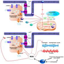
          Muscular dystrophies (MDs) are a group of inherited degenerative muscle disorders characterized by a progressive skeletal muscle wasting. Respiratory impairments and subsequent hypoxemia are encountered in a significant subgroup of patients in almost all MD forms. In response to hypoxic stress, compensatory mechanisms are activated especially through Hypoxia-Inducible Factor 1 α (HIF-1α). In healthy muscle, hypoxia and HIF-1α activation are known to affect oxidative stress balance and metabolism. Recent evidence has also highlighted HIF-1α as a regulator of myogenesis and satellite cell function. However, the impact of HIF-1α pathway modifications in MDs remains to be investigated. Multifactorial pathological mechanisms could lead to HIF-1α activation in patient skeletal muscles. In addition to the genetic defect per se, respiratory failure or blood vessel alterations could modify hypoxia response pathways. Here, we will discuss the current knowledge about the hypoxia response pathway alterations in MDs and address whether such changes could influence MD pathophysiology.

          Related collections

          Most cited references238

          • Record: found
          • Abstract: found
          • Article: not found

          Mitochondrial autophagy is an HIF-1-dependent adaptive metabolic response to hypoxia.

          Autophagy is a process by which cytoplasmic organelles can be catabolized either to remove defective structures or as a means of providing macromolecules for energy generation under conditions of nutrient starvation. In this study we demonstrate that mitochondrial autophagy is induced by hypoxia, that this process requires the hypoxia-dependent factor-1-dependent expression of BNIP3 and the constitutive expression of Beclin-1 and Atg5, and that in cells subjected to prolonged hypoxia, mitochondrial autophagy is an adaptive metabolic response which is necessary to prevent increased levels of reactive oxygen species and cell death.
            Bookmark
            • Record: found
            • Abstract: found
            • Article: found
            Is Open Access

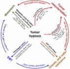
            The role of hypoxia in cancer progression, angiogenesis, metastasis, and resistance to therapy

            Hypoxia is a non-physiological level of oxygen tension, a phenomenon common in a majority of malignant tumors. Tumor-hypoxia leads to advanced but dysfunctional vascularization and acquisition of epithelial-to-mesenchymal transition phenotype resulting in cell mobility and metastasis. Hypoxia alters cancer cell metabolism and contributes to therapy resistance by inducing cell quiescence. Hypoxia stimulates a complex cell signaling network in cancer cells, including the HIF, PI3K, MAPK, and NFĸB pathways, which interact with each other causing positive and negative feedback loops and enhancing or diminishing hypoxic effects. This review provides background knowledge on the role of tumor hypoxia and the role of the HIF cell signaling involved in tumor blood vessel formation, metastasis, and development of the resistance to therapy. Better understanding of the role of hypoxia in cancer progression will open new windows for the discovery of new therapeutics targeting hypoxic tumor cells and hypoxic microenvironment.
              Bookmark
              • Record: found
              • Abstract: found
              • Article: not found

              Fiber types in mammalian skeletal muscles.

              Mammalian skeletal muscle comprises different fiber types, whose identity is first established during embryonic development by intrinsic myogenic control mechanisms and is later modulated by neural and hormonal factors. The relative proportion of the different fiber types varies strikingly between species, and in humans shows significant variability between individuals. Myosin heavy chain isoforms, whose complete inventory and expression pattern are now available, provide a useful marker for fiber types, both for the four major forms present in trunk and limb muscles and the minor forms present in head and neck muscles. However, muscle fiber diversity involves all functional muscle cell compartments, including membrane excitation, excitation-contraction coupling, contractile machinery, cytoskeleton scaffold, and energy supply systems. Variations within each compartment are limited by the need of matching fiber type properties between different compartments. Nerve activity is a major control mechanism of the fiber type profile, and multiple signaling pathways are implicated in activity-dependent changes of muscle fibers. The characterization of these pathways is raising increasing interest in clinical medicine, given the potentially beneficial effects of muscle fiber type switching in the prevention and treatment of metabolic diseases.
                Bookmark

                Author and article information

                Contributors
                Role: Academic Editor
                Journal
                Int J Mol Sci
                Int J Mol Sci
                ijms
                International Journal of Molecular Sciences
                MDPI
                1422-0067
                05 July 2021
                July 2021
                : 22
                : 13
                : 7220
                Affiliations
                [1 ]Laboratory of Respiratory Physiology, Pathophysiology and Rehabilitation, Research Institute for Health Sciences and Technology, University of Mons, 7000 Mons, Belgium; thuyhang.nguyen@ 123456umons.ac.be (T.-H.N.); stephanie.conotte@ 123456outlook.com (S.C.); alexandra.belayew@ 123456umons.ac.be (A.B.); alexandre.legrand@ 123456umons.ac.be (A.L.)
                [2 ]Department of Metabolic and Molecular Biochemistry, Research Institute for Health Sciences and Technology, University of Mons, 7000 Mons, Belgium; anne-emilie.decleves@ 123456umons.ac.be
                Author notes
                Author information
                https://orcid.org/0000-0001-5115-2424
                https://orcid.org/0000-0002-0838-2432
                https://orcid.org/0000-0002-2154-7529
                Article
                ijms-22-07220
                10.3390/ijms22137220
                8269128
                34281273
                8bbf9d19-5d86-45fd-9e93-ea1978adea3f
                © 2021 by the authors.

                Licensee MDPI, Basel, Switzerland. This article is an open access article distributed under the terms and conditions of the Creative Commons Attribution (CC BY) license ( https://creativecommons.org/licenses/by/4.0/).

                History
                : 31 May 2021
                : 30 June 2021
                Categories
                Review

                Molecular biology
                hypoxia,myopathies,hif-1α
                Molecular biology
                hypoxia, myopathies, hif-1α

                Comments

                Comment on this article