2
views
0
recommends
+1 Recommend
0 collections
    0
    shares
      • Record: found
      • Abstract: found
      • Article: found
      Is Open Access

      Pigmented median raphe cyst of the penis that developed after middle age without infection or trauma history

      case-report

      Read this article at

      Bookmark
          There is no author summary for this article yet. Authors can add summaries to their articles on ScienceOpen to make them more accessible to a non-specialist audience.

          Abstract

          Introduction

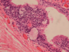
          Median raphe cysts are rare benign lesions of the male genitalia that can develop anywhere along the midline from meatus to anus. They are believed to be caused by a defect in closure of median raphe during embryonic development. These cysts commonly appear in childhood or adolescence, although some are diagnosed after middle age, typically triggered by infection or trauma. Pigmented median raphe cysts, or those containing melanin pigment and/or melanocytes, are extremely rare.

          Case presentation

          A 78‐year‐old man visited our hospital with a complaint of a penile mass that he first noticed in his 50s which slowly grew, eventually causing voiding difficulty. He had no history of infection or trauma. The lesion was excised, and the pathological diagnosis was pigmented median raphe cyst.

          Conclusion

          We successfully treated a rare case of pigmented median raphe cyst of the penis that developed after middle age without infection or trauma history.

          Related collections

          Most cited references19

          • Record: found
          • Abstract: found
          • Article: found
          Is Open Access

          Male median raphe cysts: serial retrospective analysis and histopathological classification

          Background To review the clinical and pathological characteristics of median raphe cysts and to classify the lesions according to pathogenesis and histopathological findings. Methods The medical records of patients who were diagnosed with median raphe cysts between 2001 and 2010 were reviewed to document the clinical presentation and pathological findings of the cysts. Results Most patients were asymptomatic; however, 9 patients had inflammatory or infectious cysts that were tender or painful. Four patients who had cysts on the parameatus and distal prepuce had difficulty voiding. Hematuria and hematospermia were noted in 2 cases. Thirty-one cysts were lined with an urothelium-like epithelium, and a squamous epithelium lining was found in 3 cases. In 2 cases, a well-formed mucinous glandular structure was observed. The other 20 cysts consisted of mixed epithelia. After excision of the cysts under local or general anesthesia, an urethral fistula developed as a complication in only 1 case. Conclusions Median raphe cysts are benign lesions formed due to tissue trapping during the development of urethral folds. The cysts can be defined into 4 types based on pathological findings: urethral, epidermoid, glandular, and mixed. The associated symptoms and signs should be taken into consideration when determining the treatment for the cysts. Virtual slides The virtual slide(s) for this article can be found here: http//http://www.diagnosticpathology.diagnomx.eu/vs/7727074877500751
            Bookmark
            • Record: found
            • Abstract: found
            • Article: not found

            Median raphe (parameatal) cysts of the penis.

            Although median raphe cysts of the penis have been widely reported the pathogenesis is obscure. We describe the microstructural detection of 3 cases of median raphe to study the true pathogenesis. Three patients presented to our clinic of plastic surgery complaining of abnormal shape of the penis or mass recurrence. High magnification light microscopy and electron microscopy were performed. Microstructural study revealed that all 3 cases had similar findings to those of the common urethral mucosa. Median raphe cysts are derived from the external paraurethral ducts and contain features that to our knowledge have not been previously reported.
              Bookmark
              • Record: found
              • Abstract: found
              • Article: found
              Is Open Access

              Median raphe cyst of the penis: a case report and review of the literature

              Background A defect in embryological development or closure of median raphe may lead to formation of cyst(s) anywhere in the midline from glans to anus. These cysts are referred to as median raphe cysts, an uncommonly encountered clinical condition. The cyst is generally solitary, with the penile shaft being the most common location, with average size of around 1 cm. The diagnosis is mostly clinical and confirmed histologically. We report a case of a patient with a rare histological variant of median raphe cyst and provide a focused review on presentation, histopathology, and management. Case presentation A 29-year-old unmarried Nepali man presented to our clinic with an asymptomatic, solitary, soft, translucent, nontender cystic lesion of about 1-cm diameter at the ventral aspect of glans penis, close to the meatus, that had been noticed at the age of 3 and was nonprogressive for the past 15 years. Ultrasonography demonstrated an isoechoic cystic lesion at the tip of the penis, separated from the urethra, and lying entirely within the mucosa without any evidence of solid component, septation, or vascularity. On the basis of clinical and ultrasonographic findings, a diagnosis of median raphe cyst of the penis was made. The cyst was excised with the patient under local anesthesia, and there was no evidence of recurrence in 2 years of follow-up. The histopathological examination with Hematoxylin and eosin staining showed the cyst wall was lined partly by ciliated pseudostratified columnar epithelium and partly by columnar epithelium with apical mucin. Conclusions Median raphe cyst is an uncommon, mostly asymptomatic condition in young patients. The cyst may occur anywhere along the midline from glans to anus. The diagnosis is clinical with histological confirmation. Excision is the treatment of choice with minimal chance of recurrence.
                Bookmark

                Author and article information

                Contributors
                satorutaguchi33@gmail.com
                Journal
                IJU Case Rep
                IJU Case Rep
                10.1002/(ISSN)2577-171X
                IJU5
                IJU Case Reports
                John Wiley and Sons Inc. (Hoboken )
                2577-171X
                04 March 2021
                May 2021
                : 4
                : 3 ( doiID: 10.1002/iju5.v4.3 )
                : 172-175
                Affiliations
                [ 1 ] Department of Urology Kyorin University School of Medicine Mitaka Tokyo Japan
                [ 2 ] Department of Pathology Kyorin University School of Medicine Mitaka Tokyo Japan
                [ 3 ] Department of ADPKD Research Kyorin University School of Medicine Mitaka Tokyo Japan
                Author notes
                [*] [* ] Correspondence: Satoru Taguchi M.D., Ph.D., Department of Urology, Kyorin University School of Medicine, 6‐20‐2 Shinkawa, Mitaka, Tokyo 181‐8611, Japan. Email: satorutaguchi33@ 123456gmail.com

                Author information
                https://orcid.org/0000-0002-1291-4294
                Article
                IJU512271
                10.1002/iju5.12271
                8088894
                8a26ba7c-40fa-46a6-b42c-ef6d1a85227e
                © 2021 The Authors. IJU Case Reports published by John Wiley & Sons Australia, Ltd on behalf of the Japanese Urological Association

                This is an open access article under the terms of the http://creativecommons.org/licenses/by-nc-nd/4.0/ License, which permits use and distribution in any medium, provided the original work is properly cited, the use is non‐commercial and no modifications or adaptations are made.

                History
                : 27 January 2021
                : 14 February 2021
                Page count
                Figures: 2, Tables: 1, Pages: 4, Words: 2298
                Categories
                Case Report
                Case Reports
                Custom metadata
                2.0
                May 2021
                Converter:WILEY_ML3GV2_TO_JATSPMC version:6.0.2 mode:remove_FC converted:02.05.2021

                foreskin,median raphe cyst,melanin,penis,pigmented
                foreskin, median raphe cyst, melanin, penis, pigmented

                Comments

                Comment on this article