18
views
0
recommends
+1 Recommend
0 collections
    0
    shares
      • Record: found
      • Abstract: found
      • Article: found
      Is Open Access

      Comment on outcomes in patients with esotropic Duane retraction syndrome and a partially accommodative component

      letter

      Read this article at

      Bookmark
          There is no author summary for this article yet. Authors can add summaries to their articles on ScienceOpen to make them more accessible to a non-specialist audience.

          Abstract

          Dear Sir, We read with interest the article by Kekunnaya et al.[1] The authors have conducted an interesting retrospective study and emphasized the importance of cycloplegic refraction prior to surgical management of Duane retraction syndrome (DRS) in patients with high hypermetropia. We would like to make following comments regarding their article. Cycloplegic refraction should be the primary step in the management of all patients presenting with ocular deviation. It would be disastrous to subject any child to surgery without adequate refractive correction being prescribed for an appropriate duration. 
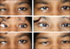
The primary cause of compensatory head posture (CHP) in DRS is limited ocular motility, with the patient adopting a posture to utilize the small field of binocular vision. The authors have not explained how elimination of head posture/torticollis occurs with spectacles. We try to explain this observation. Some children with DRS are initially able to enjoy binocular single vision (BSV) without CHP, despite motility restriction and palpebral fissure abnormality. When (later in life) the accommodative convergence induces an ocular deviation, these children probably compensate for it by adapting a CHP, which due to asymmetrical ocular motility allows them comfortable BSV. Elimination of this deviation by suitable refractive correction corrected the torticollis in these patients, probably with re-centralization of the binocular field. It is interesting to note that non-DRS patients with accommodative esodeviation cannot similarly use compensatory head posture to their advantage for BSV. In the 2nd case of Table 1, it would be interesting to know the magnitude and age at which refractive correction was prescribed. It is surprising that this child did not develop amblyopia. It would also be more informative if the authors commented about the eventual binocular status rather than simple visual acuity. Binocular functions in patients of DRS have also been controversial, and if the authors have this information about their patients, it would be a useful contribution to literature.[2] When surgical treatment is being considered, it should also be understood that angle measurements in these children are difficult and often their accuracy is doubtful. Sometimes the diagnosis is also not certain.[3] We would suggest a staged approach for their management with only medial rectus recession with or without posterior fixation being the first step. This may be combined with graded recession of ipsilateral lateral rectus if palpebral fissure abnormality is marked. 2 This would reduce the torticollis and palpebral fissure abnormality and also ensure better binocular development. More aggressive modalities like vertical rectus transposition should be taken up later in life when the measurements and results would be more predictable. The consecutive exotropia in the 2nd and 4th patient could thus be avoided.

          Related collections

          Most cited references4

          • Record: found
          • Abstract: found
          • Article: found
          Is Open Access

          Outcomes in patients with esotropic duane retraction syndrome and a partially accommodative component

          Background: The management of Duane retraction syndrome (DRS) is challenging and may become more difficult if an associated accommodative component due to high hyperopia is present. The purpose of this study is to review clinical features and outcomes in patients with partially accommodative esotropia and DRS. Setting and Design: Retrospective, non-comparative case series. Materials and Methods: Six cases of DRS with high hyperopia were reviewed. Results: Of the patients studied, the mean age of presentation was 1.3 years (range: 0.5-2.5 years). The mean amount of hyperopia was + 5D (range: 3.50-8.50) in both eyes. The mean follow up period was 7 years (range: 4 months-12 years). Five cases were unilateral while one was bilateral. Four cases underwent vertical rectus muscle transposition (VRT) and one had medial rectus recession prior to presentation; all were given optical correction. Two (50%) of the four patients who underwent vertical rectus transposition cases developed consecutive exotropia, one of whom did not have spectacles prescribed pre-operatively. All other cases (four) had minimal residual esotropia and face turn at the last follow-up with spectacle correction. Conclusion: Patients with Duane syndrome can have an accommodative component to their esotropia, which is crucial to detect and correct prior to surgery to decrease the risk of long-term over-correction. Occasionally, torticollis in Duane syndrome can be satisfactorily corrected with spectacles alone.
            Bookmark
            • Record: found
            • Abstract: not found
            • Article: not found

            Special Forms of Strabismus

              Bookmark
              • Record: found
              • Abstract: found
              • Article: found
              Is Open Access

              Congenital sixth nerve palsy or Type I Duane syndrome?

              Duane Syndrome (DS) in its classic form is characterized by congenital onset limitation of horizontal eye movements with globe retraction and narrowing of palpebral fissure (PF) on adduction.[1] Despite significant limitation of horizontal ocular motility, the ocular deviation in primary position is lesser than would occur in muscle palsies.[1] Upshoot or downshoot in adduction is commonly associated.[1] A congenital sixth nerve or congenital lateral rectus (LR) palsy is rare and may be related to birth trauma.[2] The title of a Souza-Dias publication stated: “Congenital VIth nerve is Duane's Syndrome until disproven”, and it also reflects the rarity of congenital sixth nerve paresis.[3 4] Here, we present a case of an adolescent, with a congenital sixth nerve palsy presenting as Type I DS.[5] To our knowledge, a similar case has not been reported in literature (Medline search). Our patient was successfully managed with a single muscle surgery. A 13-year-old girl presented to us with convergent strabismus in right eye (RE) since birth. Ante, peri and postnatal history were normal. There was no history of trauma or prior treatment for the strabismus. Old family photographs showed right esotropia (RET). The general and systemic examinations were essentially normal. Ocular examination revealed a visual acuity of 3/60 and N 36 in right eye which was not improving. Visual acuity in left eye was 6/6 and N 6. Refraction revealed +2 D of against the rule (ATR) HM astigmatism in RE. Prism cover test revealed an RET of 70 PD with a narrow PF RE in primary position. There was marked limitation of abduction of RE with widening of the PF on abduction. Adduction of RE was slightly limited with upshoot and further narrowing of PF. The esotropia had a “V” pattern of 20 PD. There was no compensatory head posture [Figure 1]. There were no binocular functions on Worth Four Dot and Randot Stereo acuity Tests. Forced duction testing revealed a tight medial rectus RE. The patient underwent 5 mm recession of right medial rectus (MR) by conjunctival limbal approach and standard technique.[6] A very tight MR muscle was encountered intraoperatively with thin blue sclera under MR muscle. The patient was followed up on day 1, 7, 14 and 60 postoperatively. Postoperative examinations revealed RET of 10 PD in primary position. Ocular movements showed no change in abduction. Compared to preoperative adduction movement, a greater decrease in RE was noted. The abnormality in PF size in different gazes including the primary position had reduced markedly. The residual V pattern was of 12 PD. The patient was satisfied with the cosmesis [Figures 2 and 3]. Figure 1 Preoperative photographs. Note the palpebral fissure abnormality and “V” pattern of esodevation Figure 2 Postoperative photographs. Note the correction of deviation in primary position, correction of “V” pattern Figure 3 Preoperative (on left) and postoperative (on right) photographs in primary position. Note the correction of palpebral fissure abnormality An esotropic DS is more common than a congenital sixth nerve palsy. The findings of ET in primary position with restricted abduction and associated narrowing of PF in adduction in our patient are consistent with Type I DS.[1 5] Slight limitation of adduction with upshoot is also found in DS.[1 3] However, the primary position ET is relatively small in DS (less than 30 PD) compared to LR pasly or paresis.[7] Although widening of PF in abduction is typical of DS,[1 3] PF narrowing is not a very dependable diagnostic sign of mild to moderate DS[3] as narrowing of the PF on adduction is usually interpreted as a passive adjustment of the lids to retracting globe.[1] Our patient was diagnosed as a case of congenital sixth nerve palsy, with ocular motility, PF abnormalities, and forced duction test results mimicking Type I DS. The nature of abduction in upgaze to downgaze of LR palsy is a V pattern, compared with the curved outward rotation (X pattern) uniquely characteristic of DS[8] [Figure 4]. The presence of V pattern in our patient and absence of anomalous LR innervations in upgaze and downgaze points toward an LR palsy rather than a DS. The alteration in PF size can be explained by a secondary contracture of RMR muscle due to a longstanding LR palsy.[3] The tight MR would cause retraction of the globe on adduction with consequent narrowing of PF. It also explains the marginally limited adduction and upshoot.[3] MR would relax in abduction, hence the apparent PF widening. The correction of PF abnormality and limitation of adduction postoperatively also support the presence of a fibrotic MR muscle. The presence of thin blue sclera under MR confirms the direction of MR pressure indentation.[3] On the other hand, MR in children with DS does not exhibit excessive stiffness or contracture in the primary zone, that is, it is normal.[9] Figure 4 Diagrammatic representation of ocular deviation in dextroelevation, dextroversion and dextrodepression in the presence of (a) Duane Syndrome (b) infantile esotropia and (c) congenital sixth nerve palsy in the right eye. Note the “X” pattern of deviation in (a), a straight line (no incomitance) in (b) and “V” pattern in (c). An “A” or “V” pattern may be seen in infantile esotropia, but it is not apparent when the involved eye is in abduction Infantile esotropia also forms a differential diagnosis of this presentation, but it usually has full ductions. A diagnosis of congenital LR palsy with tight MR was made on the basis of the above features. The collapse of V pattern can be attributed to correction of deviation in primary position. A V pattern of up to 15 PD (residual V of 12 PD in our patient) is considered physiological.[10] On further retrospection, correction of such a large esodeviation in DS would have required a more aggressive surgery like asymmetric MR recessions or transpositions of the SR/IR muscles temporally.[11–14] We could have probably avoided the postoperative adduction deficit in MR by reducing the amount of MR recession or by putting the tight muscle on hang back sutures.
                Bookmark

                Author and article information

                Journal
                Indian J Ophthalmol
                Indian J Ophthalmol
                IJO
                Indian Journal of Ophthalmology
                Medknow Publications & Media Pvt Ltd (India )
                0301-4738
                1998-3689
                August 2014
                : 62
                : 8
                : 895
                Affiliations
                [1]Department of Ophthalmology, King Georges’ Medical University, Lucknow, Uttar Pradesh, India
                Author notes
                Correspondence to: Dr. Siddharth Agrawal, Department of Ophthalmology, King Georges’ Medical University, Lucknow - 226 003, Uttar Pradesh, India. E-mail: agrawalsiddharth@ 123456rediffmail.com
                Article
                IJO-62-895a
                10.4103/0301-4738.141069
                4185174
                25230971
                8a2691ed-947d-40a7-a530-887bcc37976e
                Copyright: © Indian Journal of Ophthalmology

                This is an open-access article distributed under the terms of the Creative Commons Attribution-Noncommercial-Share Alike 3.0 Unported, which permits unrestricted use, distribution, and reproduction in any medium, provided the original work is properly cited.

                History
                Categories
                Letters to the Editor

                Ophthalmology & Optometry
                Ophthalmology & Optometry

                Comments

                Comment on this article