- Record: found
- Abstract: found
- Article: found
Congenital sixth nerve palsy or Type I Duane syndrome?
other
Read this article at
There is no author summary for this article yet. Authors can add summaries to their articles on ScienceOpen to make them more accessible to a non-specialist audience.
Abstract
Duane Syndrome (DS) in its classic form is characterized by congenital onset limitation
of horizontal eye movements with globe retraction and narrowing of palpebral fissure
(PF) on adduction.[1] Despite significant limitation of horizontal ocular motility,
the ocular deviation in primary position is lesser than would occur in muscle palsies.[1]
Upshoot or downshoot in adduction is commonly associated.[1] A congenital sixth nerve
or congenital lateral rectus (LR) palsy is rare and may be related to birth trauma.[2]
The title of a Souza-Dias publication stated: “Congenital VIth nerve is Duane's Syndrome
until disproven”, and it also reflects the rarity of congenital sixth nerve paresis.[3
4] Here, we present a case of an adolescent, with a congenital sixth nerve palsy presenting
as Type I DS.[5] To our knowledge, a similar case has not been reported in literature
(Medline search). Our patient was successfully managed with a single muscle surgery.
A 13-year-old girl presented to us with convergent strabismus in right eye (RE) since
birth. Ante, peri and postnatal history were normal. There was no history of trauma
or prior treatment for the strabismus. Old family photographs showed right esotropia
(RET). The general and systemic examinations were essentially normal. Ocular examination
revealed a visual acuity of 3/60 and N 36 in right eye which was not improving. Visual
acuity in left eye was 6/6 and N 6. Refraction revealed +2 D of against the rule (ATR)
HM astigmatism in RE. Prism cover test revealed an RET of 70 PD with a narrow PF RE
in primary position. There was marked limitation of abduction of RE with widening
of the PF on abduction. Adduction of RE was slightly limited with upshoot and further
narrowing of PF. The esotropia had a “V” pattern of 20 PD. There was no compensatory
head posture [Figure 1]. There were no binocular functions on Worth Four Dot and Randot
Stereo acuity Tests. Forced duction testing revealed a tight medial rectus RE. The
patient underwent 5 mm recession of right medial rectus (MR) by conjunctival limbal
approach and standard technique.[6] A very tight MR muscle was encountered intraoperatively
with thin blue sclera under MR muscle. The patient was followed up on day 1, 7, 14
and 60 postoperatively. Postoperative examinations revealed RET of 10 PD in primary
position. Ocular movements showed no change in abduction. Compared to preoperative
adduction movement, a greater decrease in RE was noted. The abnormality in PF size
in different gazes including the primary position had reduced markedly. The residual
V pattern was of 12 PD. The patient was satisfied with the cosmesis [Figures 2 and
3].
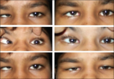
Figure 1
Preoperative photographs. Note the palpebral fissure abnormality and “V” pattern of
esodevation
Figure 2
Postoperative photographs. Note the correction of deviation in primary position, correction
of “V” pattern
Figure 3
Preoperative (on left) and postoperative (on right) photographs in primary position.
Note the correction of palpebral fissure abnormality
An esotropic DS is more common than a congenital sixth nerve palsy. The findings of
ET in primary position with restricted abduction and associated narrowing of PF in
adduction in our patient are consistent with Type I DS.[1
5] Slight limitation of adduction with upshoot is also found in DS.[1
3] However, the primary position ET is relatively small in DS (less than 30 PD) compared
to LR pasly or paresis.[7]
Although widening of PF in abduction is typical of DS,[1
3] PF narrowing is not a very dependable diagnostic sign of mild to moderate DS[3]
as narrowing of the PF on adduction is usually interpreted as a passive adjustment
of the lids to retracting globe.[1]
Our patient was diagnosed as a case of congenital sixth nerve palsy, with ocular motility,
PF abnormalities, and forced duction test results mimicking Type I DS. The nature
of abduction in upgaze to downgaze of LR palsy is a V pattern, compared with the curved
outward rotation (X pattern) uniquely characteristic of DS[8] [Figure 4]. The presence
of V pattern in our patient and absence of anomalous LR innervations in upgaze and
downgaze points toward an LR palsy rather than a DS. The alteration in PF size can
be explained by a secondary contracture of RMR muscle due to a longstanding LR palsy.[3]
The tight MR would cause retraction of the globe on adduction with consequent narrowing
of PF. It also explains the marginally limited adduction and upshoot.[3] MR would
relax in abduction, hence the apparent PF widening. The correction of PF abnormality
and limitation of adduction postoperatively also support the presence of a fibrotic
MR muscle. The presence of thin blue sclera under MR confirms the direction of MR
pressure indentation.[3] On the other hand, MR in children with DS does not exhibit
excessive stiffness or contracture in the primary zone, that is, it is normal.[9]
Figure 4
Diagrammatic representation of ocular deviation in dextroelevation, dextroversion
and dextrodepression in the presence of (a) Duane Syndrome (b) infantile esotropia
and (c) congenital sixth nerve palsy in the right eye. Note the “X” pattern of deviation
in (a), a straight line (no incomitance) in (b) and “V” pattern in (c). An “A” or
“V” pattern may be seen in infantile esotropia, but it is not apparent when the involved
eye is in abduction
Infantile esotropia also forms a differential diagnosis of this presentation, but
it usually has full ductions. A diagnosis of congenital LR palsy with tight MR was
made on the basis of the above features.
The collapse of V pattern can be attributed to correction of deviation in primary
position. A V pattern of up to 15 PD (residual V of 12 PD in our patient) is considered
physiological.[10] On further retrospection, correction of such a large esodeviation
in DS would have required a more aggressive surgery like asymmetric MR recessions
or transpositions of the SR/IR muscles temporally.[11–14] We could have probably avoided
the postoperative adduction deficit in MR by reducing the amount of MR recession or
by putting the tight muscle on hang back sutures.
Related collections
Most cited references16
- Record: found
- Abstract: not found
- Article: not found
Electrophysiology of the retraction syndromes.
Lukas Huber (1974)
- Record: found
- Abstract: found
- Article: not found
Vertical rectus muscle transposition surgery for Duane's syndrome.
A Molarte, A Rosenbaum (2015)
- Record: found
- Abstract: found
- Article: not found
Effect of transposition surgery on rectus muscle paths by magnetic resonance imaging.
J Demer, Robert Miller, Miriam A. Rosenbaum (1993)