1
views
0
recommends
+1 Recommend
0 collections
    0
    shares
      • Record: found
      • Abstract: found
      • Article: found
      Is Open Access

      Stigmasterol alleviates interleukin-1beta-induced chondrocyte injury by down-regulatingsterol regulatory element binding transcription factor 2 to regulateferroptosis

      research-article
      , ,
      Bioengineered
      Taylor & Francis
      Stigmasterol, knee osteoarthritis, chondrocytes, ferroptosis

      Read this article at

      Bookmark
          There is no author summary for this article yet. Authors can add summaries to their articles on ScienceOpen to make them more accessible to a non-specialist audience.

          ABSTRACT

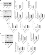
          Stigmasterol (STM), one of the main active components of Achyranthes bidentata, has been shown to effectively inhibit proinflammatory factors and matrix degradation in chondrocytes. However, the effect of STM on interleukin (IL)-1β-induced chondrocytes and its specific mechanism remain unclear. The purpose of the present study was to explore the effect and mechanism of sterol regulatory element binding transcription factor 2 (SREBF2) on IL-1β induced chondrocytes in the presence of STM. CCK-8 was used to detect the effect of STM on the cell viability of mouse chondrogenic cells (ATDC5). After ATDC5 cells were induced by IL-1β, the expression of SREBF2 in osteoarthritis cells was detected by RT-qPCR. The content of iron ion in the cells was detected by using an iron colorimetric assay kit. After further transfection of a SREBF2 overexpressing vector (Oe-SREBF2) or addition of a ferroptosis inhibitor, the expression levels of inflammation and matrix degradation-related proteins were detected via Western blotting. The levels of oxidative stress in cells were determined by using an ELISA kit. The results revealed that STM had no significant effect on the viability of ATDC5 cells. STM reduced IL-1β-induced ATDC5 cell damage and ferroptosis through SREBF2 and enhanced the inhibitory effect of ferroptosis inhibitors on IL-1β-induced ATDC5 cell injury. The present data suggest that STM attenuated chondrocyte injury induced by IL-1β by regulating ferroptosis via down-regulation of SREBF2, and may have potential as a novel therapeutic method for knee osteoarthritis.

          Related collections

          Most cited references30

          • Record: found
          • Abstract: found
          • Article: not found

          Analysis of relative gene expression data using real-time quantitative PCR and the 2(-Delta Delta C(T)) Method.

          The two most commonly used methods to analyze data from real-time, quantitative PCR experiments are absolute quantification and relative quantification. Absolute quantification determines the input copy number, usually by relating the PCR signal to a standard curve. Relative quantification relates the PCR signal of the target transcript in a treatment group to that of another sample such as an untreated control. The 2(-Delta Delta C(T)) method is a convenient way to analyze the relative changes in gene expression from real-time quantitative PCR experiments. The purpose of this report is to present the derivation, assumptions, and applications of the 2(-Delta Delta C(T)) method. In addition, we present the derivation and applications of two variations of the 2(-Delta Delta C(T)) method that may be useful in the analysis of real-time, quantitative PCR data. Copyright 2001 Elsevier Science (USA).
            Bookmark
            • Record: found
            • Abstract: found
            • Article: not found

            Ferroptosis: A Regulated Cell Death Nexus Linking Metabolism, Redox Biology, and Disease

            Ferroptosis is a form of regulated cell death characterized by the iron-dependent accumulation of lipid hydroperoxides to lethal levels. Emerging evidence suggests that ferroptosis represents an ancient vulnerability caused by the incorporation of polyunsaturated fatty acids into cellular membranes, and cells have developed complex systems that exploit and defend against this vulnerability in different contexts. The sensitivity to ferroptosis is tightly linked to numerous biological processes, including amino acid, iron, and polyunsaturated fatty acid metabolism, and the biosynthesis of glutathione, phospholipids, NADPH, and coenzyme Q10. Ferroptosis has been implicated in the pathological cell death associated with degenerative diseases (i.e., Alzheimer's, Huntington's, and Parkinson's diseases), carcinogenesis, stroke, intracerebral hemorrhage, traumatic brain injury, ischemia-reperfusion injury, and kidney degeneration in mammals and is also implicated in heat stress in plants. Ferroptosis may also have a tumor-suppressor function that could be harnessed for cancer therapy. This Primer reviews the mechanisms underlying ferroptosis, highlights connections to other areas of biology and medicine, and recommends tools and guidelines for studying this emerging form of regulated cell death.
              Bookmark
              • Record: found
              • Abstract: found
              • Article: not found

              Reactive Oxygen Species, Aging and Articular Cartilage Homeostasis

              Chondrocytes are responsible for the maintenance of the articular cartilage. A loss of homeostasis in cartilage contributes to the development of osteoarthritis (OA) when the synthetic capacity of chondrocytes is overwhelmed by processes that promote matrix degradation. There is evidence for an age-related imbalance in reactive oxygen species (ROS) production relative to the anti-oxidant capacity of chondrocytes that plays a role in cartilage degradation as well as chondrocyte cell death. The ROS produced by chondrocytes that have received the most attention include superoxide, hydrogen peroxide, the reactive nitrogen species nitric oxide, and the nitric oxide derived product peroxynitrite. Excess levels of these ROS not only cause oxidative-damage but, perhaps more importantly, cause a disruption in cell signaling pathways that are redox-regulated, including Akt and MAP kinase signaling. Age-related mitochondrial dysfunction and reduced activity of the mitochondrial superoxide dismutase (SOD2) are associated with an increase in mitochondrial-derived ROS and are in part responsible for the increase in chondrocyte ROS with age. Peroxiredoxins (Prxs) are a key family of peroxidases responsible for removal of H2O2, as well as for regulating redox-signaling events. Prxs are inactivated by hyperoxidation. An age-related increase in chondrocyte Prx hyperoxidation and an increase in OA cartilage has been noted. The finding in mice that deletion of SOD2 or the anti-oxidant gene transcriptional regulator nuclear factor-erythroid 2- related factor (Nrf2) result in more severe OA, while overexpression or treatment with mitochondrial targeted anti-oxidants reduces OA, further support a role for excessive ROS in the pathogenesis of OA. Therefore, new therapeutic strategies targeting specific anti-oxidant systems including mitochondrial ROS may be of value in reducing the progression of age-related OA.
                Bookmark

                Author and article information

                Journal
                Bioengineered
                Bioengineered
                Bioengineered
                Taylor & Francis
                2165-5979
                2165-5987
                22 November 2021
                2021
                22 November 2021
                : 12
                : 2
                : 9332-9340
                Affiliations
                [0001]Department of Orthopedics, Shenzhen Hospital of Beijing University of Chinese Medicine (Longgang); , Shenzhen, P.R. China
                Author notes
                CONTACT Zhisheng Mo mozhisheng8100@ 123456163.com Shenzhen Hospital of Beijing University of Chinese Medicine (Longgang); , 1 Dayun Road, Longcheng Street, Longgang District, Shenzhen, Guangdong 518100, P.R. China
                Article
                2000742
                10.1080/21655979.2021.2000742
                8810005
                34806937
                884f23aa-12cf-4221-9e8b-ccc8fe0deeb3
                © 2021 The Author(s). Published by Informa UK Limited, trading as Taylor & Francis Group.

                This is an Open Access article distributed under the terms of the Creative Commons Attribution License ( http://creativecommons.org/licenses/by/4.0/), which permits unrestricted use, distribution, and reproduction in any medium, provided the original work is properly cited.

                History
                Page count
                Figures: 4, References: 30, Pages: 9
                Categories
                Research Article
                Research Paper

                Biomedical engineering
                stigmasterol,knee osteoarthritis,chondrocytes,ferroptosis
                Biomedical engineering
                stigmasterol, knee osteoarthritis, chondrocytes, ferroptosis

                Comments

                Comment on this article