7
views
0
recommends
+1 Recommend
0 collections
    0
    shares
      • Record: found
      • Abstract: found
      • Article: found
      Is Open Access

      Low-intensity pulsed ultrasound improves behavioral and histological outcomes after experimental traumatic brain injury

      research-article
      1 , 2 , 3 , 4 , , 1 , 5 ,
      Scientific Reports
      Nature Publishing Group UK

      Read this article at

      Bookmark
          There is no author summary for this article yet. Authors can add summaries to their articles on ScienceOpen to make them more accessible to a non-specialist audience.

          Abstract

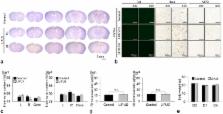
          The purpose of this study was to investigate the neuroprotective effects of low-intensity pulsed ultrasound (LIPUS) on behavioral and histological outcomes in a mouse model of traumatic brain injury (TBI). Mice subjected to controlled cortical impact injury were treated with LIPUS in the injured region daily for a period of 4 weeks. The effects of LIPUS on edema were observed by MR imaging in the mouse brain at 1 and 4 days following TBI. Brain water content, blood-brain barrier permeability, histology analysis, and behavioral studies were performed to assess the effects of LIPUS. Two-way analysis of variance and Student t test were used for statistical analyses, with a significant level of 0.05. Treatment with LIPUS significantly attenuated brain edema, blood-brain barrier permeability, and neuronal degeneration beginning at day 1. Compared with the TBI group, LIPUS also significantly improved functional recovery and reduced contusion volumes up to post-injury day 28. Post-injury LIPUS treatment reduced brain edema and improved behavioral and histological outcomes following TBI. The neuroprotective effects of LIPUS may be a promising new technique for treating TBI.

          Related collections

          Most cited references30

          • Record: found
          • Abstract: found
          • Article: not found

          Inflammatory response in acute traumatic brain injury: a double-edged sword.

          Inflammation is an important part of the pathophysiology of traumatic brain injury. Although the central nervous system differs from the other organs because of the almost complete isolation from the blood stream mediated by the blood-brain barrier, the main steps characterizing the immune activation within the brain follow a scenario similar to that in other organs. The key players in these processes are the numerous immune mediators released within minutes of the primary injury. They guide a sequence of events including expression of adhesion molecules, cellular infiltration, and additional secretion of inflammatory molecules and growth factors, resulting in either regeneration or cell death. The question is this: to what extent is inflammation beneficial for the injured brain tissue, and how does it contribute to secondary brain damage and progressive neuronal loss? This review briefly reports recent evidence supporting the dual, the beneficial, or the deleterious role of neuroinflammation after traumatic brain injury.
            Bookmark
            • Record: found
            • Abstract: found
            • Article: not found

            Clinical trials in head injury.

            Traumatic brain injury (TBI) remains a major public health problem globally. In the United States the incidence of closed head injuries admitted to hospitals is conservatively estimated to be 200 per 100,000 population, and the incidence of penetrating head injury is estimated to be 12 per 100,000, the highest of any developed country in the world. This yields an approximate number of 500,000 new cases each year, a sizeable proportion of which demonstrate significant long-term disabilities. Unfortunately, there is a paucity of proven therapies for this disease. For a variety of reasons, clinical trials for this condition have been difficult to design and perform. Despite promising pre-clinical data, most of the trials that have been performed in recent years have failed to demonstrate any significant improvement in outcomes. The reasons for these failures have not always been apparent and any insights gained were not always shared. It was therefore feared that we were running the risk of repeating our mistakes. Recognizing the importance of TBI, the National Institute of Neurological Disorders and Stroke (NINDS) sponsored a workshop that brought together experts from clinical, research, and pharmaceutical backgrounds. This workshop proved to be very informative and yielded many insights into previous and future TBI trials. This paper is an attempt to summarize the key points made at the workshop. It is hoped that these lessons will enhance the planning and design of future efforts in this important field of research.
              Bookmark
              • Record: found
              • Abstract: found
              • Article: not found

              Progressive atrophy and neuron death for one year following brain trauma in the rat.

              Although atrophic changes have been well described following traumatic brain injury (TBI) in humans, little is known concerning the mechanisms or progression of brain tissue loss. In the present study, we evaluated the temporal profile of histopathological changes following parasagittal fluid-percussion (FP) brain injury in rats over 1 year postinjury. Anesthetized 3-4 month-old Sprague-Dawley Rats (n = 51) were subjected to FP brain injury of high severity (2.5-2.9 atm, n = 51) or sham treatment (n = 27). At 1 h, 2 h, 48 h, 1 week, 2 weeks, 1 month, 2 months, 6 months and 1 year after brain injury or sham treatment, these animals were humanely euthanized. Brain sections were analyzed with image-processing techniques to determine the extent of cortical tissue loss and shrinkage of the hippocampal pyramidal cell layer. In addition, cell counting was performed to determine the number of neurons in the dentate hilus of the hippocampus, and glial fibrillary acidic protein (GFAP) immunostaining was used to reveal reactive astrocytosis. Examination of the injured brains revealed substantial and progressive tissue loss with concomitant ventriculomegaly in the hemisphere ipsilateral to injury. The regions with the most notable progressive atrophy included the cortex, hippocampus, thalamus, and septum. Quantitative analysis demonstrated a significantly progressive loss of cortical tissue as well as shrinkage of the hippocampal pyramidal cell layer ipsilateral to injury over 1 year following injury. In addition, reactive astrocytosis in regions of atrophy and progressive bilateral death of neurons in the dentate hilus was observed for 1 year following injury. These results suggest that a chronically progressive degenerative process may be initiated by brain trauma. Thus, there is a temporally broad window within which to introduce novel therapeutic strategies designed to ameliorate the short and long-term consequences of brain trauma.
                Bookmark

                Author and article information

                Contributors
                szufuchen@yahoo.com.tw
                fyyang@ym.edu.tw
                Journal
                Sci Rep
                Sci Rep
                Scientific Reports
                Nature Publishing Group UK (London )
                2045-2322
                14 November 2017
                14 November 2017
                2017
                : 7
                : 15524
                Affiliations
                [1 ]ISNI 0000 0001 0425 5914, GRID grid.260770.4, Department of Biomedical Imaging and Radiological Sciences, National Yang-Ming University, ; Taipei, Taiwan
                [2 ]ISNI 0000 0004 0634 0356, GRID grid.260565.2, Graduate Institute of Life Sciences, National Defense Medical Center, ; Taipei, Taiwan
                [3 ]ISNI 0000 0004 0634 0356, GRID grid.260565.2, Departments of Physiology and Biophysics, National Defense Medical Center, ; Taipei, Taiwan
                [4 ]ISNI 0000 0004 0572 7890, GRID grid.413846.c, Department of Physical Medicine and Rehabilitation, Cheng Hsin General Hospital, ; Taipei, Taiwan
                [5 ]ISNI 0000 0001 0425 5914, GRID grid.260770.4, Biophotonics and Molecular Imaging Research Center, National Yang-Ming University, ; Taipei, Taiwan
                Article
                15916
                10.1038/s41598-017-15916-2
                5686128
                29138458
                84902208-c9ba-4913-ab0a-b464c0b2f5ee
                © The Author(s) 2017

                Open Access This article is licensed under a Creative Commons Attribution 4.0 International License, which permits use, sharing, adaptation, distribution and reproduction in any medium or format, as long as you give appropriate credit to the original author(s) and the source, provide a link to the Creative Commons license, and indicate if changes were made. The images or other third party material in this article are included in the article’s Creative Commons license, unless indicated otherwise in a credit line to the material. If material is not included in the article’s Creative Commons license and your intended use is not permitted by statutory regulation or exceeds the permitted use, you will need to obtain permission directly from the copyright holder. To view a copy of this license, visit http://creativecommons.org/licenses/by/4.0/.

                History
                : 14 June 2017
                : 3 November 2017
                Categories
                Article
                Custom metadata
                © The Author(s) 2017

                Uncategorized
                Uncategorized

                Comments

                Comment on this article